Volume 10, Number 10—October 2004
Dispatch
Fatal Naegleria fowleri Meningoencephalitis, Italy
Paola E. Cogo* , Massimo Scaglia†, Simonetta Gatti†, Flavio Rossetti‡, Rita Alaggio*, Anna Maria Laverda*, Ling Zhou§, Lihua Xiao§, and Govinda S. Visvesvara§
Figure 1
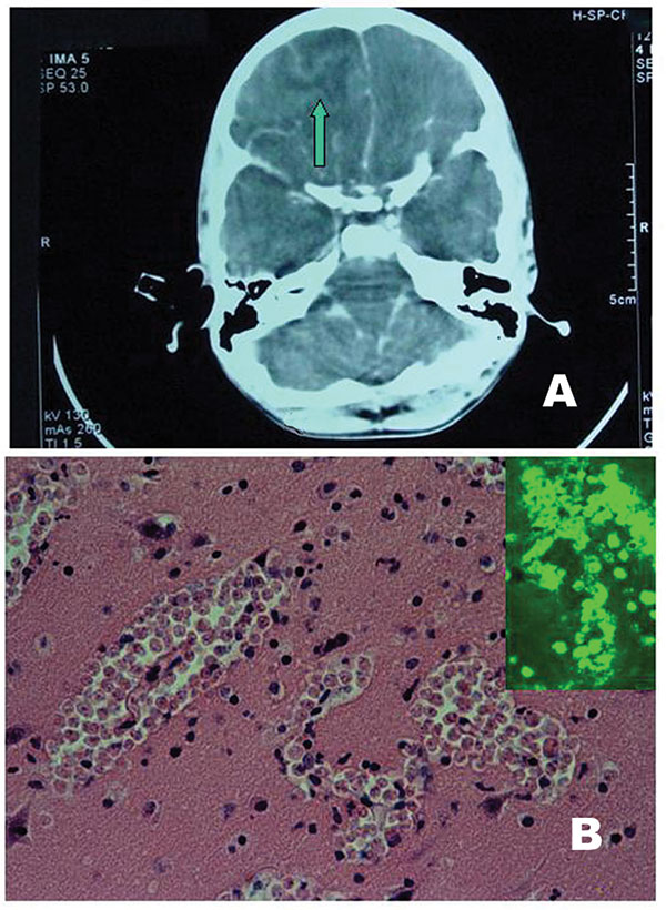
Figure 1. A) Computed tomographic scan: note the right fronto-basal collection (arrow) with a midline shift right to left. B) Brain histology: three large clusters of amebic vegetative forms are seen (H-E stain, x 250). Inset: Positive indirect immunofluorescent analysis on tissue section with anti– Naegleria fowleri serum.
Page created: April 11, 2011
Page updated: April 11, 2011
Page reviewed: April 11, 2011
The conclusions, findings, and opinions expressed by authors contributing to this journal do not necessarily reflect the official position of the U.S. Department of Health and Human Services, the Public Health Service, the Centers for Disease Control and Prevention, or the authors' affiliated institutions. Use of trade names is for identification only and does not imply endorsement by any of the groups named above.