Volume 10, Number 11—November 2004
Research
Topographic Changes in SARS Coronavirus–infected Cells during Late Stages of Infection
Mah-Lee Ng* , J.W.M. Lee*, M.L.N. Leong*, A.-E. Ling†, H.-C. Tan‡, and E.E. Ooi‡
Figure 2
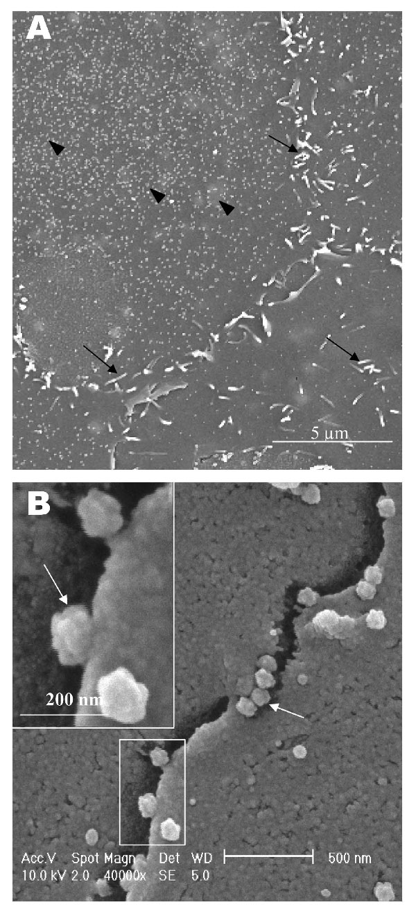
Figure 2. Scanning electron microscopy of Vero E6 cells infected with severe acute respiratory syndrome–associated coronavirus at 15 h after infection. A) One pronounced surface morphologic change is the proliferation of psuedopodia at the cell periphery (arrows). Some pseudopodia are also developing on the cell surface. Some cells appear to have large amount of extracellular virus on the cell surface (arrowhead), whereas neighboring cells seem deprived of any extracelluar virus particles. B) Virus particles are protruding from the edge of cells (arrows). Inset shows the boxed area at higher magnification. Virus particles appear knobby and rosettelike.
Page created: April 21, 2011
Page updated: April 21, 2011
Page reviewed: April 21, 2011
The conclusions, findings, and opinions expressed by authors contributing to this journal do not necessarily reflect the official position of the U.S. Department of Health and Human Services, the Public Health Service, the Centers for Disease Control and Prevention, or the authors' affiliated institutions. Use of trade names is for identification only and does not imply endorsement by any of the groups named above.