Volume 10, Number 11—November 2004
Research
Topographic Changes in SARS Coronavirus–infected Cells during Late Stages of Infection
Mah-Lee Ng* , J.W.M. Lee*, M.L.N. Leong*, A.-E. Ling†, H.-C. Tan‡, and E.E. Ooi‡
Figure 3
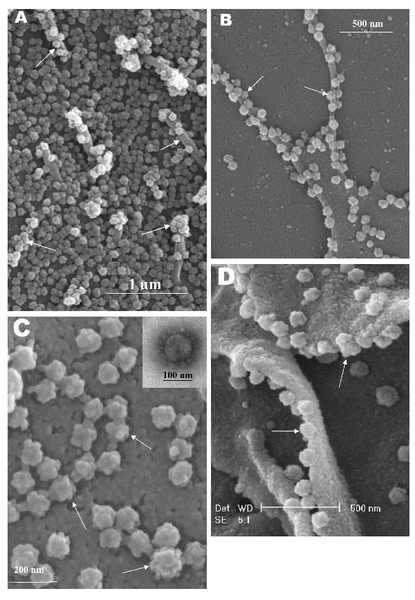
Figure 3. Scanning electron microscopy of Vero E6 cells infected with severe acute respiratory syndrome–associated coronavirus at 24 h after infection. A) Cell surface is covered with extracellular progeny virus particles, and progeny virus are being extruded from or attached to numerous pseudopodia on infected cell surface (arrows). B) A higher magnification micrograph of the virus-clustered pseudopodia (arrows). C) Rosettelike appearance of the matured virus particles (arrows). The scanning electron microscopy image complements the form and structure of the virus seen with negative staining (inset) under transmission electron microscopy. Short and stubby spikes are visible on the virus surface.
Page created: April 21, 2011
Page updated: April 21, 2011
Page reviewed: April 21, 2011
The conclusions, findings, and opinions expressed by authors contributing to this journal do not necessarily reflect the official position of the U.S. Department of Health and Human Services, the Public Health Service, the Centers for Disease Control and Prevention, or the authors' affiliated institutions. Use of trade names is for identification only and does not imply endorsement by any of the groups named above.