Skip directly to site content Skip directly to page options Skip directly to A-Z link Skip directly to A-Z link Skip directly to A-Z link
Volume 10, Number 2—February 2004
THEME ISSUE
2004 SARS Edition
Laboratory Study

Ultrastructural Characterization of SARS Coronavirus

Cynthia S. Goldsmith*, Kathleen M. Tatti*, Thomas G. Ksiazek*, Pierre E. Rollin*, James A. Comer*, William W. Lee*, Paul A. Rota*, Bettina Bankamp*, William J. Bellini*, and Sherif R. Zaki*
Author affiliations: *Centers for Disease Control and Prevention, Atlanta, Georgia, USA

Main Article

Figure 1

Assembly of severe acute respiratory syndrome–associated coronavirus (SARS-CoV) particles in infected Vero E6 cells. A) Apposition of nucleocapsids (arrow) along membranes of the budding compartment as particles developed and budded. Nucleocapsids measured 6 nm in diameter and were mostly seen in cross-section. Some virions had an electron-lucent center, with the nucleocapsid juxtaposed to the envelope, while others were relatively dark when the nucleocapsid was present throughout the particle.

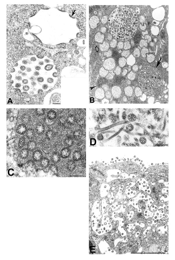
Figure 1. Assembly of severe acute respiratory syndrome–associated coronavirus (SARS-CoV) particles in infected Vero E6 cells. A) Apposition of nucleocapsids (arrow) along membranes of the budding compartment as particles developed and budded. Nucleocapsids measured 6 nm in diameter and were mostly seen in cross-section. Some virions had an electron-lucent center, with the nucleocapsid juxtaposed to the envelope, while others were relatively dark when the nucleocapsid was present throughout the particle. Tannic acid pre-treatment enhanced the visibility of the club-shaped viral projections (inset), which averaged 14 nm in length. B) SARS-CoV–infected cell with virus-containing vesicles, double-membrane vesicles (open arrow), and nucleocapsid inclusions (arrowhead). Note the vesicle with granular material interspersed among the virions (arrow). C) Higher magnification of a virus-containing vesicle with dark granular material. D) Tubular structures in a virus-containing vesicle. E) Virions in vesicles, which appeared to migrate toward and fuse with the plasma membrane. The characteristic lining of particles along the cell surface is seen. Bars: A, inset; B–D, 100 nm; E, 1 μm.

Main Article

Page created: January 31, 2011
Page updated: January 31, 2011
Page reviewed: January 31, 2011
The conclusions, findings, and opinions expressed by authors contributing to this journal do not necessarily reflect the official position of the U.S. Department of Health and Human Services, the Public Health Service, the Centers for Disease Control and Prevention, or the authors' affiliated institutions. Use of trade names is for identification only and does not imply endorsement by any of the groups named above.
file_external