Volume 10, Number 3—March 2004
Research
Amoebae-resisting Bacteria Isolated from Human Nasal Swabs by Amoebal Coculture
Gilbert Greub*1 , Bernard La Scola*, and Didier Raoult*
Figure 2
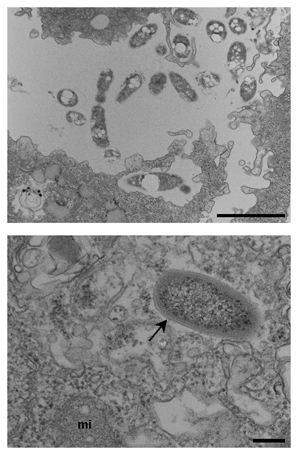
Figure 2. A. Bosea sp. (isolate 3) in the process of being phagocytized by Acanthamoeba polyphaga (arrows) and in the extracellular media, as seen on electron microscopy. 8,900 x magnification. Bar represents 200 nm. B. Chryseobacterium-like rod (isolate 7) within A. polyphaga (arrows), as seen on electron microscopy. Arrow show the trilammelar membrane. mi = amoebal mitochondria. 36,000 x magnification. Bar represents 200 nm.
1G. Greub’s current affiliation is University of Lausanne, Lausanne, Switzerland.
Page created: June 14, 2011
Page updated: June 14, 2011
Page reviewed: June 14, 2011
The conclusions, findings, and opinions expressed by authors contributing to this journal do not necessarily reflect the official position of the U.S. Department of Health and Human Services, the Public Health Service, the Centers for Disease Control and Prevention, or the authors' affiliated institutions. Use of trade names is for identification only and does not imply endorsement by any of the groups named above.