Volume 11, Number 10—October 2005
Dispatch
Diphyllobothriasis, Brazil
Jorge Luiz Mello Sampaio* , Victor Piana de Andrade*, Maria da Conceição Lucas*, Liang Fung*, Sandra Maria B. Gagliardi*, Sandra Rosalem P. Santos*, Caio Marcio Figueiredo Mendes*, Maria Bernadete de Paula Eduardo†, and Terry Dick‡
Figure
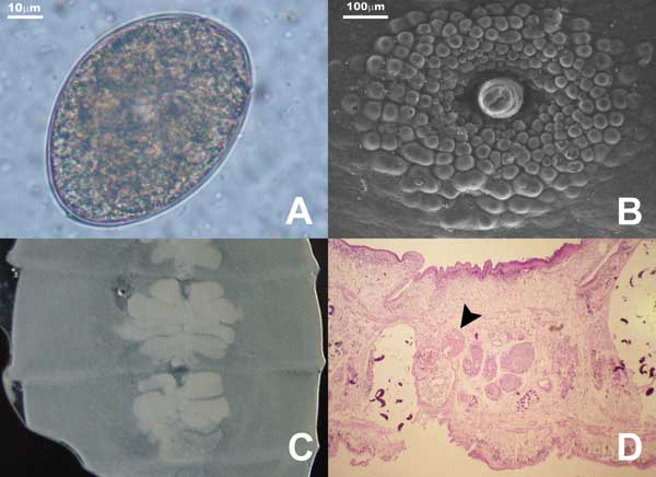
Figure. A) Diphyllobothrium latum egg. Note opercular constriction. B) Genital papillae of mature proglottids as seen under scanning electron microscope. C) Uterine loops of gravid proglottids in fresh preparation. D) Sagittal section of the genital pore region stained with hematoxylin-eosin. Note seminal vesicle (arrowhead) situated dorsocaudal to the cirrus sac (magnification 100×).
Page created: February 22, 2012
Page updated: February 22, 2012
Page reviewed: February 22, 2012
The conclusions, findings, and opinions expressed by authors contributing to this journal do not necessarily reflect the official position of the U.S. Department of Health and Human Services, the Public Health Service, the Centers for Disease Control and Prevention, or the authors' affiliated institutions. Use of trade names is for identification only and does not imply endorsement by any of the groups named above.