Volume 11, Number 3—March 2005
Dispatch
Mimivirus in Pneumonia Patients
Bernard La Scola*, Thomas J. Marrie†, Jean-Pierre Auffray‡, and Didier Raoult*
Figure
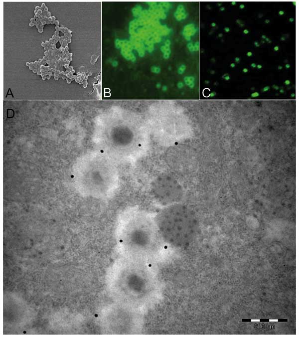
Figure. . As observed by scanning electronic microscopy, mimivirus antigen (A) is recognized by antibodies in our microimmunofluorescence assay using conventional fluorescence microscope (B) and confocal microscope (C). Mature particles within amebas are also recognized by antibodies seen with transmission electronic microscopy immunogold technique (D) (mimivirus particle size 400 nm).
Page created: April 25, 2012
Page updated: April 25, 2012
Page reviewed: April 25, 2012
The conclusions, findings, and opinions expressed by authors contributing to this journal do not necessarily reflect the official position of the U.S. Department of Health and Human Services, the Public Health Service, the Centers for Disease Control and Prevention, or the authors' affiliated institutions. Use of trade names is for identification only and does not imply endorsement by any of the groups named above.