Skip directly to site content Skip directly to page options Skip directly to A-Z link Skip directly to A-Z link Skip directly to A-Z link
Volume 12, Number 12—December 2006
Dispatch

West Nile Virus in Horses, sub-Saharan Africa

Olivier Cabre*Comments to Author , Marc Grandadam†, Jean-Lou Marié‡, Patrick Gravier†, Aurélie Prangé†, Yan Santinelli§, Vincent Rous¶, Olivier Bourry#, Jean-Paul Durand†, Hugues J. Tolou†, and Bernard Davoust**
Author affiliations: *École du Val-de-Grâce, Paris, France; †Institut de Médecine Tropicale du Service de Santé des Armées, Marseille, France; ‡Secteur Vétérinaire de Marseille, Marseille, France; §Service Vétérinaire du Régiment de Cavalerie de la Garde Républicaine, Paris, France; ¶Secteur Vétérinaire de Lyon, Lyon, France; #Centre International de Recherches Médicales, Franceville, Gabon; **Direction Régionale du Service de Santé des Armées de Toulon, Toulon, France

Main Article

Figure 2

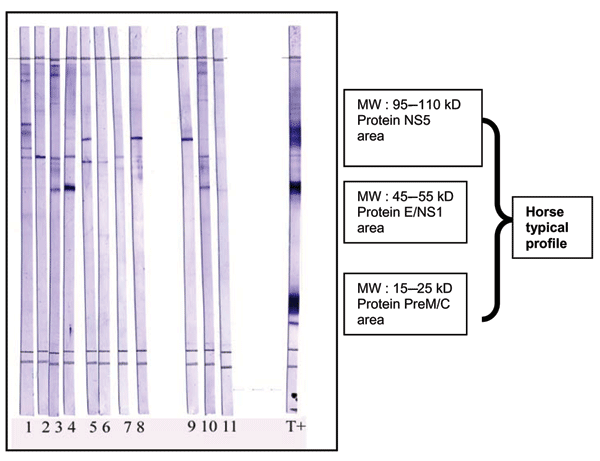
West Nile virus (WNV) Western blot as a validation of ELISA-positive results. Proteins from WNV-infected cells lysates were separated by sodium dodecyl sulfate–polyacrylamide gel electrophoresis on 13% acrylamide gels and transferred on polyvinylidene difluoride membranes. Strips were cut and blocked with nonfat milk (5%). Serum samples diluted (1:200) in phosphate-buffered saline (PBS), 5% nonfat milk, and 0.1% Tween 20 were loaded on strips and incubated for 1 h with slow shaking. Strips were

Figure 2. West Nile virus (WNV) Western blot as a validation of ELISA-positive results. Proteins from WNV-infected cells lysates were separated by sodium dodecyl sulfate–polyacrylamide gel electrophoresis on 13% acrylamide gels and transferred on polyvinylidene difluoride membranes. Strips were cut and blocked with nonfat milk (5%). Serum samples diluted (1:200) in phosphate-buffered saline (PBS), 5% nonfat milk, and 0.1% Tween 20 were loaded on strips and incubated for 1 h with slow shaking. Strips were washed 4 times in PBS, 0.5% Tween, and incubated for 1 h in anti-horse immunoglobulin G (IgG) peroxidase (1:8,000). After 4 washes, Western blots were incubated for 5 min with trimethylbenzidine substrate (with specific enhancer). Strips were washed once with water to stop the staining. Results were obtained within 24 h. MW, molecular weight; T+, horse from Chad with positive Western blot results; serum samples 1–11, horse from southeast France, IgG positive by ELISA but negative by neutralization; NS5, nonstructural protein 5; E, envelope; PreM, premembrane; C, capsid.

Main Article

Page created: October 04, 2011
Page updated: October 04, 2011
Page reviewed: October 04, 2011
The conclusions, findings, and opinions expressed by authors contributing to this journal do not necessarily reflect the official position of the U.S. Department of Health and Human Services, the Public Health Service, the Centers for Disease Control and Prevention, or the authors' affiliated institutions. Use of trade names is for identification only and does not imply endorsement by any of the groups named above.
file_external