Volume 12, Number 2—February 2006
Dispatch
Evaluation of a Direct, Rapid Immunohistochemical Test for Rabies Diagnosis
Figure 2
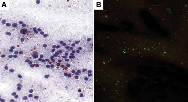
Figure 2. Touch impression of a deteriorated glycerolated brain from a Tanzanian spotted hyena (Crocuta crocuta) with rabies. A) Brain processed by direct rapid immunohistochemical test (dRIT). Magnification, ×400. B) DFA staining procedure on the same brain. Magnification, ×200.
Page created: February 02, 2012
Page updated: February 02, 2012
Page reviewed: February 02, 2012
The conclusions, findings, and opinions expressed by authors contributing to this journal do not necessarily reflect the official position of the U.S. Department of Health and Human Services, the Public Health Service, the Centers for Disease Control and Prevention, or the authors' affiliated institutions. Use of trade names is for identification only and does not imply endorsement by any of the groups named above.