Volume 12, Number 4—April 2006
Synopsis
Domestic Ducks and H5N1 Influenza Epidemic, Thailand
Thaweesak Songserm*, Rungroj Jam-on*, Numdee Sae-Heng*, Noppadol Meemak†, Diane J. Hulse-Post‡, Katharine Sturm-Ramirez‡, and Robert G. Webster‡
Figure 4
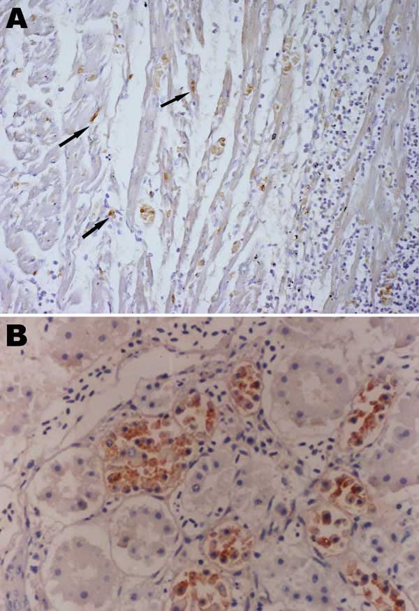
Figure 4. Immunohistochemistry of an HPAI H5N1–infected white Cherry Valley duck (Anas platyrhynchos). The viral antigen is detected in myocardial cells and lymphoid cells (arrow) (A) and renal tubular cells (B) (magnification ×100). The primary antibody used for immunohistochemistry in this study was a mouse anti–avian influenza H5 antibody (Magellan Biotechnology, Chunan, Taiwan).
Page created: January 23, 2012
Page updated: January 23, 2012
Page reviewed: January 23, 2012
The conclusions, findings, and opinions expressed by authors contributing to this journal do not necessarily reflect the official position of the U.S. Department of Health and Human Services, the Public Health Service, the Centers for Disease Control and Prevention, or the authors' affiliated institutions. Use of trade names is for identification only and does not imply endorsement by any of the groups named above.