Volume 12, Number 9—September 2006
Research
Histologic Features and Immunodetection of African Tick-bite Fever Eschar
Hubert Lepidi*, Pierre-Edouard Fournier*, and Didier Raoult*
Figure 4
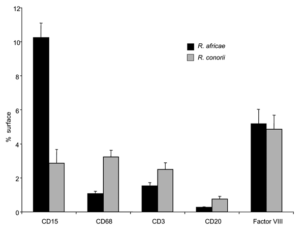
Figure 4. Quantification of inflammatory changes in inoculation eschars from patients with African tick-bite fever (Rickettsia africae, n = 8) and patients with Mediterranean spotted fever (R. conorii, n = 15). Surface areas expressing CD15, CD68, CD3, CD20, and Factor VIII were quantified after immunostaining. Quantification of each parameter was evaluated by computer-assisted analysis of digitized microscopic images. Results were normalized and expressed as a percentage of the total skin tissue surface area. Columns represent mean values ± standard error.
Page created: November 17, 2011
Page updated: November 17, 2011
Page reviewed: November 17, 2011
The conclusions, findings, and opinions expressed by authors contributing to this journal do not necessarily reflect the official position of the U.S. Department of Health and Human Services, the Public Health Service, the Centers for Disease Control and Prevention, or the authors' affiliated institutions. Use of trade names is for identification only and does not imply endorsement by any of the groups named above.