Volume 14, Number 1—January 2008
Research
Experimental Infection of Swans and Geese with Highly Pathogenic Avian Influenza Virus (H5N1) of Asian Lineage
Justin D. Brown* , David E. Stallknecht*, and David E. Swayne†
Figure 1
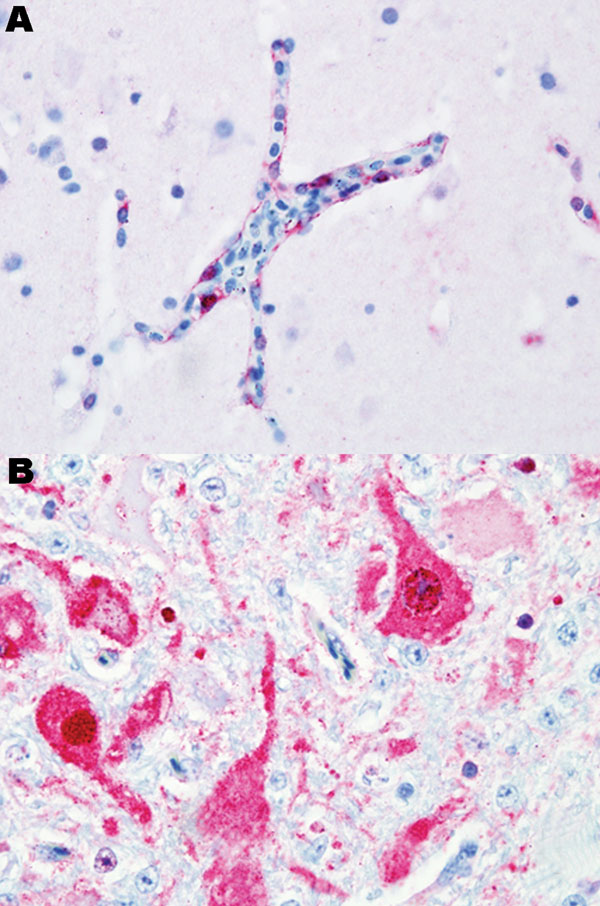
Figure 1. Photomicrograph of viral antigen (red). A) Endothelial cells lining a blood vessel in the brain of a black swan. B) Neurons in the brain of a mute swan. Both birds died after experimental infection with highly pathogenic avian influenza virus (H5N1). Immunohistochemical stain with hematoxylin counterstain. Magnification ×40.
Page created: July 08, 2010
Page updated: July 08, 2010
Page reviewed: July 08, 2010
The conclusions, findings, and opinions expressed by authors contributing to this journal do not necessarily reflect the official position of the U.S. Department of Health and Human Services, the Public Health Service, the Centers for Disease Control and Prevention, or the authors' affiliated institutions. Use of trade names is for identification only and does not imply endorsement by any of the groups named above.