Volume 15, Number 2—February 2009
Dispatch
Mycobacterium bolletii Respiratory Infections
Abstract
Contrary to other species in the Mycobacterium chelonae-abscessus complex, we reidentified M. bolletii strains isolated from 4 respiratory patients and found these strains to be uniformly resistant to clarithromycin. No mutations previously associated with macrolide resistance in bacteria were detected in either the 23S rDNA or the genes encoding riboproteins L4 and L22.
Mycobacterium chelonae-abscessus complex (MCAC) members are opportunistic pathogens in patients with underlying pulmonary disorders (1–3). We recently described M. bolletii as a new MCAC member (4); the pathogen was later isolated from 9% of cystic fibrosis patients (5). The initial description of M. bolletii suggested that it was highly resistant to antimicrobial drugs, including clarithromycin (4). To gain a better appreciation of this resistance, we reidentified MCAC isolates collected in our microbiology laboratory, Timone Hospital, Marseilles, during the past 10 years and performed in vitro susceptibility testing and sequencing of the 23S rDNA, L4, and L22 genes.
Patient 1, a 47-year-old woman with an unremarkable medical history, sought treatment after a 3-week history of hemoptysis; radiographs showed bilateral micronodular infiltrates in the upper lung lobes. Blood tests showed a polymorphonuclear cell count of 9 × 10³/mL and biological inflammatory syndrome. Bleeding in the left lobar bronchus was observed during bronchoscopy. Microbiologic results of a bronchial lavage specimen were negative, and microscopic examination found no acid-fast bacilli. An isolate from a subsequent sputum sample was identified later as M. bolletii, however. The patient was discharged. No further information is available for this patient.
Patient 2, a 76-year-old man who had received treatment for pulmonary tuberculosis (TB) in 1976, sought treatment in November 1995 for hemoptysis and signs and symptoms of broncho-pulmonary infection. A chest radiograph showed a large cavity in the right upper lobe and infiltrate in the left upper lobe. Two stomach aspirates yielded an isolate identified as M. abscessus despite negative results of a direct microscopic examination; refined identification performed 8 years later in 2003 found both isolates to be M. bolletii. The patient received rifampin, clarithromycin, isoniazid, and ciprofloxacin for 16 months. After an initial improvement, the patient continued to have an episodic cough and hemoptysis, and M. bolletii was grown from 2 sputum specimens; direct microscopic examination yielded acid-fast bacilli. In November 1998, the patient still had symptoms. In 2003, he was admitted to an intensive care unit for acute respiratory distress syndrome. A broncho-alveolar lavage specimen yielded a mixed culture of multidrug-resistant M. bolletii and Klebsiella pneumoniae despite negative results of direct examination. The patient eventually died of K. pneumoniae septicemia within days of his admission to the ICU.
Patient 3, a 77-year-old man who smoked 3 packs of cigarettes per month and who had a history of pulmonary TB, was admitted to Timone Hospital, Marseilles, in October 2000 with a diagnosis of bronchitis. Corticoid therapy was prescribed. In March 2001, the patient was admitted for respiratory insufficiency. A chest radiograph showed diffuse bullous emphysema in both lungs. Microscopic examination of 1 sputum specimen and 1 bronchial aspirate yielded the presence of acid-fast bacilli that were later identified as M. bolletii. The patient left the hospital without treatment, and no further information on his condition is available.
Patient 4, a 90-year-old woman, sought treatment with a temperature of 38°C, hemoptysis, and bilateral micronodular infiltrates of the upper lung lobes. She had a history of childhood pulmonary TB. Sputum specimens yielded mycobacteria that were identified later as M. bolletii despite negative results of direct examination. The patient remained febrile after 1 month of treatment with intravenous imipenem and amikacin, her pulmonary condition worsened, and a sputum smear showed numerous acid-fast bacilli. The treatment regimen was changed to a combination of ciprofloxacin, clarithromycin, and ethambutol, but the patient died 2 weeks after treatment began.
Thirty-one isolates, previously identified as M. abscessus (n = 20) and M. chelonae (n = 11) by 16S rDNA sequencing from January 1996 through June 2007, were reidentified by partial rpoB gene sequencing as described (6). The MICs of 22 antimicrobial drugs were determined by E-test (AB Biodisk, Solna, Sweden) after the culture was incubated for 3 days at 30°C. Data were interpreted by using the broth microdilution criteria (7). A 1,500-bp fragment of the 23S rDNA (8), a 635-bp fragment of the L4 ribosomal protein (forward primer L4Absc7F: 5′-ATCGCAGTCAAGGCTCCGG-3′, reverse primer L4Absc642R: 5′-TCAGGCCGACACCTCCTC-3′), and a 471-bp fragment of the L22 ribosomal protein (forward primer L22Absc1F: 5′-ATGACCACTACTACCGAAT-3′, reverse primer L22Absc471R: 5′-CTAGCTGGTGCCTCCCTT-3′) were PCR amplified and sequenced from clarithromycin-resistant isolates. The study was approved by local Ethics Committee, Marseilles Medical School.
Figure
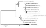
Figure. Phylogenetic tree of different sequevars of Mycobacterium abscessus-chelonnae group isolates determined by rpoB-723 bp sequencing. Isolate identified as M. bolletii is in boldface. Scale bar represents 1% sequence divergence.
Twelve of 31 isolates were identified by rpoB sequencing as M. abscessus, 11 as M. chelonae, 4 as M. massiliense, and 4 as M. bolletii. M. abscessus and M. bolletii isolates showed no intraspecific rpoB sequence variation. In contrast, 0.7% sequence divergence was observed in M. chelonae (6 sequevars) and M. massiliense (3 sequevars) isolates (Figure). The 4 M. bolletii isolates were unique among these 31 MCAC isolates in that they were multidrug resistant (Table). The isolates exhibited clarithromycin MICs >256 µg/mL, whereas the other MCAC isolates had clarithromycin MICs <2 µg/mL. The E-test is not a validated method for MIC determination in rapidly growing mycobacteria, yet the results we obtained were similar to those previously reported for the reference broth microdilution method (9). In the M. bolletii isolates, we found no substitutions, deletions, or insertions in domain V (A2058, A2059, C2611 position, Escherichia coli numbering) of the 23S rDNA or in the L4 and L22 ribosomal protein genes.
M. bolletii is an emerging pathogen responsible for respiratory tract infections in patients with underlying compromised respiratory function. In our study, M. bolletii was responsible for pulmonary infection in 3 of 4 patients (patients 2–4) (10). M. bolletii was repeatedly isolated from different samples from those 3 patients over a period of several weeks. A coexisting broncho-pulmonary disease was found in 3 of the 4 patients, and clinical features and radiograph patterns that suggested nontuberculous mycobacteria infection were observed in all 4 patients (10). Infected patients were >75 years of age. Three of 4 patients had hemoptysis and lung infiltrates, M. bolletii was the sole organism isolated from respiratory tract specimens. In this study, 16S rDNA sequencing misidentified 25% of 31 MCAC isolates that were eventually identified as M. massiliense and M. bolletii. This statistic agrees with observations made during the recent description of M. bolletii infection after mesotherapy (11).
Clarithromycin was administrated to 2 of the 4 patients, both of whom died within several weeks after treatment began. The 4 M. bolletii isolates were highly resistant to clarithromycin, yet they did not harbor the 23S rDNA mutations that have been previously found in clarithromycin-resistant M. abscessus strains (12). Additionally, no mutations in riboproteins L4 and L22, which are associated with macrolide resistance in Streptococcus pneumoniae (13), were detected. RNA methylase genes erm[38], erm[39], and erm[40], which confer inducible macrolide resistance in M. fortuitum, M. smegmatis, M. mageritense, and M. wolinskyi, are absent in the MCAC (14). Further investigations are therefore needed to clarify the mechanism of clarithromycin resistance in M. bolletii.
Clarithromycin has been recommended as the first-line antimicrobial drug for treating rapidly growing mycobacteria infections in patients with compromised respiratory function (1,10). Patient deaths (3) have been linked to clarithromycin resistance, with a risk of secondary clarithromycin resistance during monotherapy estimated to be <10% (12). Recent studies showed that 21%–36% of MCAC isolates were resistant to clarithromycin (15). The recommendation for treating M. abscessus infection with clarithromycin was made before discovering M. bolletii, a multidrug-resistant species that mimics M. abscessus. Our report illustrates that accurate species identification and in vitro clarithromycin susceptibility testing should be recommended for MCAC isolates of clinical interest. GenBank accession nos. were as follows: for 23S rDNA sequences, M. bolletii CIP 108541T (EU109306), M. massiliense CIP 108297T (EU109307), M. abscessus CIP 104536T (EU109308), and M. chelonae CIP 104535T (EU109309); for L4 sequences, M. bolletii CIP 108541T M. massiliense (EU779956), CIP 108297T (EU779957), M. abscessus CIP 104536T (EU779957), and M. chelonae CIP 104535T (EU779958); and for L22 sequences M. bolletii CIP 108541T (EU779952), M. massiliense CIP 108297T (EU779953), M. abscessus CIP 104536T (EU779954), and M. chelonae CIP 104535T (could not be amplified with the designed primers).
Dr Adékambi is an American Society for Microbiology postdoctoral fellow at the Mycobacteriology Laboratory Branch, Centers for Diseases Control and Prevention, Atlanta, Georgia. His research interests include detection, identification, and characterization of mycobacteria in clinical and environmental samples.
Dr Drancourt is professor of microbiology at Marseille Medical School, Marseille, France. His research interests are the molecular identification of bacteria such as mycobacteria, as well as paleomicrobiology.
Acknowledgments
We thank Christian de Fontaine for technical assistance.
This study was supported by Oeuvre Antituberculeuse des Bouches du Rhône, Marseille, France.
References
- Brown-Elliott BA, Wallace RJ Jr. Clinical and taxonomic status of pathogenic nonpigmented or late-pigmenting rapidly growing mycobacteria. Clin Microbiol Rev. 2002;15:716–46. DOIPubMedGoogle Scholar
- Griffith DE. Emergence of nontuberculous mycobacteria as pathogens in cystic fibrosis. Am J Respir Crit Care Med. 2003;167:810–2. DOIPubMedGoogle Scholar
- Sanguinetti M, Ardito F, Fiscarelli E, La Sorda M, D’Argenio P, Ricciotti G, Fatal pulmonary infection due to multidrug-resistant Mycobacterium abscessus in a patient with cystic fibrosis. J Clin Microbiol. 2001;39:816–9. DOIPubMedGoogle Scholar
- Adekambi T, Berger P, Raoult D, Drancourt M. rpoB gene sequence-based characterization of emerging non-tuberculous mycobacteria with descriptions of Mycobacterium bolletii sp. nov., Mycobacterium phocaicum sp. nov. and Mycobacterium aubagnense sp. nov. Int J Syst Evol Microbiol. 2006;56:133–43. DOIPubMedGoogle Scholar
- Roux AL, Catherinot E, Rippoll F, Soismier N, Guterriez C, Vincent V, Mycobacteries non-tuberculeuses et mucoviscidoses: enquête française de prevalence. Abstract 9ème Colloque des Jeunes Chercheurs en Mucoviscidose, Paris, 2008.
- Adékambi T, Colson P, Drancourt M. rpoB-based identification of nonpigmented and late-pigmenting rapidly growing mycobacteria. J Clin Microbiol. 2003;41:5699–708. DOIPubMedGoogle Scholar
- National Committee for Clinical Laboratory Standards. Susceptibility testing of Mycobacteria, Nocardia, and other aerobic actinomycetes. Approved standard M24-A. Wayne (PA): The Committee; 2003.
- Meier A, Kirschner P, Springer B, Steingrube VA, Brown BA, Wallace RJ Jr, Identification of mutations in 23S rRNA gene of clarithromycin-resistant Mycobacterium intracellulare. Antimicrob Agents Chemother. 1994;38:381–4.PubMedGoogle Scholar
- Simmon KE, Pounder JI, Greene JN, Walsh F, Anderson CM, Cohen S, Identification of an emerging pathogen, Mycobacterium massiliense, by rpoB sequencing of clinical isolates collected in the United States. J Clin Microbiol. 2007;45:1978–80. DOIPubMedGoogle Scholar
- Griffith DE, Aksamit T, Brown-Elliott BA, Catanzaro A, Daley C, Gordin F, An official ATS/IDSA statement: diagnosis, treatment, and revention of nontuberculous mycobacterial diseases. Am J Respir Crit Care Med. 2007;175:367–416. DOIPubMedGoogle Scholar
- Viana-Niero C, Lima KV, Lopes ML, Rabello MC, Marsola LR, Brilhante VC, Molecular characterization of Mycobacterium massiliense and Mycobacterium bolletii in isolates collected from outbreaks of infections after laparoscopic surgeries and cosmetic procedures. J Clin Microbiol. 2008;46:850–5. DOIPubMedGoogle Scholar
- Wallace RJ Jr, Meier A, Brown BA, Zhang Y, Sander P, Onyi GO, Genetic basis for clarithromycin resistance among isolates of Mycobacterium chelonae and Mycobacterium abscessus. Antimicrob Agents Chemother. 1996;40:1676–81.PubMedGoogle Scholar
- Canu A, Malbruny B, Coquemont M, Davies TA, Appelbaum PC, Leclercq R. Diversity of ribosomal mutations conferring resistance to macrolides, clindamycin, streptogramin, and telithromycin in Streptococcus pneumoniae. Antimicrob Agents Chemother. 2002;46:125–31. DOIPubMedGoogle Scholar
- Nash KA, Andini N, Zhang Y, Brown-Elliott BA, Wallace RJ Jr. Intrinsic macrolide resistance in rapidly growing mycobacteria. Antimicrob Agents Chemother. 2006;50:3476–8. DOIPubMedGoogle Scholar
- Yang SC, Hsueh PR, Lai HC, Teng LJ, Huang LM, Chen JM, High prevalence of antimicrobial resistance in rapidly growing mycobacteria in Taiwan. Antimicrob Agents Chemother. 2003;47:1958–62. DOIPubMedGoogle Scholar
Figure
Table
Cite This ArticleTable of Contents – Volume 15, Number 2—February 2009
| EID Search Options |
|---|
|
|
|
|
|
|
Please use the form below to submit correspondence to the authors or contact them at the following address:
Michel Drancourt, Unité des Rickettsies, Faculté de Médecine, 27 Blvd Jean Moulin, 13385 Marseille CEDEX 5, France
Top