Volume 15, Number 7—July 2009
Letter
Ranavirus Outbreak in North American Bullfrogs (Rana catesbeiana), Japan, 2008
Yumi Une , Akiko Sakuma, Hiroki Matsueda, Katsuki Nakai, and Masaru Murakami
Appendix Figure
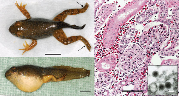
Appendix Figure. North American bullfrog (Rana catesbeiana) metamorphs infected with ranavirus RCV-JP. A) Necrosis of distal extremities (arrows) and mild abdominal swelling. Scale bar = 1 cm. B) Severe abdominal swelling caused by body cavity effusion. Scale bar = 1 cm. C) Kidney of an infected frog with necrosis of glomeruli and tubular hyaline droplet degeneration; hematoxylin and eosin stain. Scale bar = 100 m. Inset shows ranavirus-like particles; scale bar = 100 nm.
Page created: November 09, 2010
Page updated: November 09, 2010
Page reviewed: November 09, 2010
The conclusions, findings, and opinions expressed by authors contributing to this journal do not necessarily reflect the official position of the U.S. Department of Health and Human Services, the Public Health Service, the Centers for Disease Control and Prevention, or the authors' affiliated institutions. Use of trade names is for identification only and does not imply endorsement by any of the groups named above.