Volume 15, Number 9—September 2009
Research
Susceptibilities of Nonhuman Primates to Chronic Wasting Disease
Figure 2
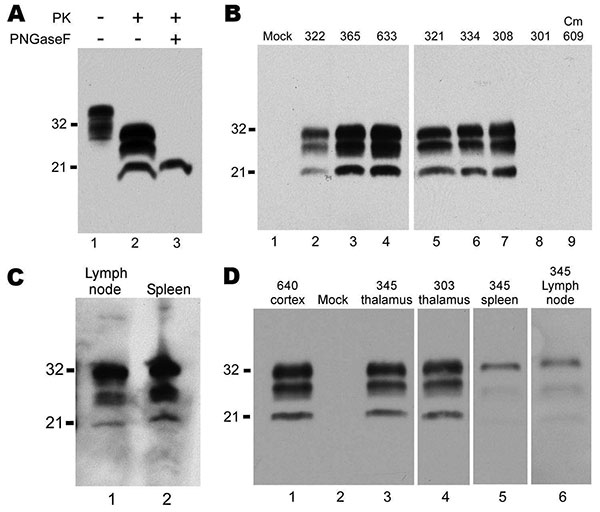
Figure 2. Western blots of squirrel monkey protease-resistant prion protein (PrPres). A) Brain homogenate from squirrel monkey 322, showing proteinase K (PK)–resistant PrPres. A downward shift of 7–9 kDa after PK digestion indicated a banding pattern typical of PrPres (lane 2). After deglycosylation with peptide-N-glycosidase F, 1 band of PrPres was present (lane 3). Lane 1, 0.5 mg of brain tissue equivalents; lane 2, 0.6 mg; lane 3, 0.4 mg. Blot was developed by using antibody 3F4 against PrP, enhanced chemiluminescence (ECL), and a 2-min exposure. B) Brains of monkeys screened for PrPres. All tissues were treated with PK, and lanes were loaded with 0.25 mg brain tissue equivalents, except for lane 5, which was loaded with 1.0 mg. All lanes contain samples of brain cortex except lanes 6 and 7, which contain thalamus. Blot was developed by using antibody 3F4, ECL, and a 30-min exposure. Lane 1, control. In lanes 2–7, PrP banding is similar among squirrel monkeys infected with different pools of chronic wasting disease (CWD) agent (Table 1). PrPres was not detected in brain of orally infected squirrel monkey 301 (lane 8) or in brain of an intracerebrally infected cynomolgus macaque (Cm) 609 (lane 9). C) Lymphatic tissues from squirrel monkey 365. For visualization of PrPres in lymph node and spleen, increased amounts of tissue were loaded, and a more sensitive detection system (femto detection; Thermo Scientific, Waltham, MA, USA) was used. Lane 1, lymph node, 0.7 mg tissue equivalents; lane 2, spleen, 1.1 mg tissue equivalents. Blot was developed by using antibody 3F4, femto-enhanced ECL, and a 1-min exposure. D) PK-treated brain and lymphatic tissues from orally infected squirrel monkeys 303 and 345. Lane 1, positive control no. 640; lane 2, negative control; lane 3, no. 345 thalamus; lane 4, no. 303 thalamus; lane 5, no. 345 spleen; lane 6, no. 345 mesenteric lymph node. Bands were visualized by using antibody 3F4 (residues 109–112) and ECL. Lanes 1–5, 10-min exposure; lane 6, overnight exposure; tissue equivalents loaded per lane: lanes 1–4, 0.25 mg; lane 5, 0.5 mg; lane 6, 1 mg. Values on the left of all blots are in kDa.