Volume 17, Number 1—January 2011
Dispatch
Serodiagnosis of Primary Infections with Human Parvovirus 4, Finland
Anne Lahtinen, Pia Kivelä, Lea Hedman, Arun Kumar, Anu Kantele, Maija Lappalainen, Kirsi Liitsola, Matti Ristola, Eric Delwart, Colin P. Sharp, Peter Simmonds, Maria Söderlund-Venermo, and Klaus Hedman
Figure 1
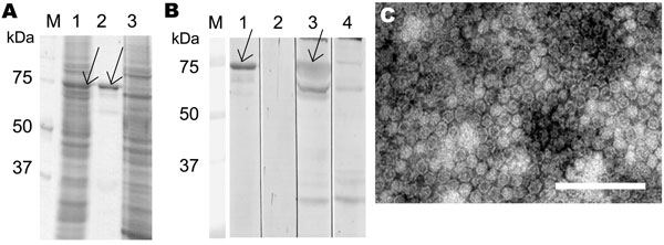
Figure 1. Parvovirus 4 (PARV4) virus-like particle (VLP) expression and immunoreactivity, Finland. A) Sodium dodecyl sulfate–polyacrylamide gel electrophoresis of PARV4-like particles in Spodoptera frugiperda armyworm (Sf)9 cells (lane 1), purified VLPs (lane 2), and uninfected Sf9 cells (lane 3). B) Western blotting with PARV4 immunoglobulin (Ig) G–positive serum (lanes 1, 3, and 4) or PARV4 IgG–negative serum (lane 2). Lanes 1 and 2, purified VLPs as antigen; lane 3, Sf9 cells expressing VLPs; lane 4, Sf9 cells expressing glutathione-S-transferase control antigen; lanes M, molecular mass marker. Arrows in panels A and B indicate the PARV4 capsid protein. C) Electron micrograph of purified VLPs. Scale bar = 200 nm.
Page created: July 08, 2011
Page updated: July 08, 2011
Page reviewed: July 08, 2011
The conclusions, findings, and opinions expressed by authors contributing to this journal do not necessarily reflect the official position of the U.S. Department of Health and Human Services, the Public Health Service, the Centers for Disease Control and Prevention, or the authors' affiliated institutions. Use of trade names is for identification only and does not imply endorsement by any of the groups named above.