Volume 17, Number 2—February 2011
Dispatch
Blastomycosis in Man after Kinkajou Bite
Julie R. Harris , David D. Blaney, Mark D. Lindsley, Sherif R. Zaki, Christopher D. Paddock, Clifton P. Drew, April J. Johnson, Douglas Landau, Joel Vanderbush, and Robert Baker
Figure 1
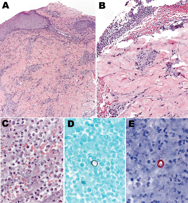
Figure 1. Histologic appearance of the cutaneous lesion of a man with blastomycosis. Ulcerated epidermis (A) showing superficial and deep perivascular infiltrates, predominantly mononuclear inflammatory cells. Fibrinopurulent exudate (B) adjacent to the ulcer, comprising neutrophils, erythrocytes, and necrotic cellular debris (C), and occasional large yeasts morphologically compatible with Blastomyces dermatitidis infection (D and E). Hematoxylin and eosin stain (A, B, and C), Grocott methenamine silver stain (D), and immunoalkaline phosphatase with antibody against B. dermatitidis and naphthol fast red with hematoxylin counterstain (E). Original magnifications ×12.5 (A), ×25 (B), and ×100 (C–E).
Page created: July 13, 2011
Page updated: July 13, 2011
Page reviewed: July 13, 2011
The conclusions, findings, and opinions expressed by authors contributing to this journal do not necessarily reflect the official position of the U.S. Department of Health and Human Services, the Public Health Service, the Centers for Disease Control and Prevention, or the authors' affiliated institutions. Use of trade names is for identification only and does not imply endorsement by any of the groups named above.