Volume 17, Number 5—May 2011
Dispatch
Linguatula serrata Tongue Worm in Human Eye, Austria
Martina Koehsler, Julia Walochnik , Michael Georgopoulos, Christian Pruente, Wolfgang Boeckeler, Herbert Auer, and Talin Barisani-Asenbauer
Figure 1
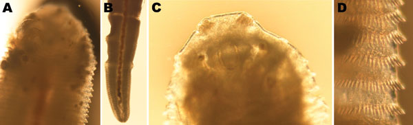
Figure 1. Morphology of Linguatula serrata tongue worm. A) Ventral anterior end with hooks. B) Posterior end laterally with primordial uterus, genital porus, and intestine (note peristalsis). C) Dorsal anterior end with cuticular structures of apical papillae, chitinoid oral clasp, and insertions of oral muscles. D) Rows of spicules. Original magnification ×50 (A–C) or ×100 (D).
Page created: August 02, 2011
Page updated: August 02, 2011
Page reviewed: August 02, 2011
The conclusions, findings, and opinions expressed by authors contributing to this journal do not necessarily reflect the official position of the U.S. Department of Health and Human Services, the Public Health Service, the Centers for Disease Control and Prevention, or the authors' affiliated institutions. Use of trade names is for identification only and does not imply endorsement by any of the groups named above.