Volume 19, Number 1—January 2013
Dispatch
Sheep-to-Human Transmission of Orf Virus during Eid al-Adha Religious Practices, France
Antoine Nougairede1 , Christelle Fossati1, Nicolas Salez, Stephan Cohen-Bacrie, Laetitia Ninove, Fabrice Michel, Samer Aboukais, Mathias Buttner, Christine Zandotti, Xavier de Lamballerie, and Remi N. Charrel
Figure 1
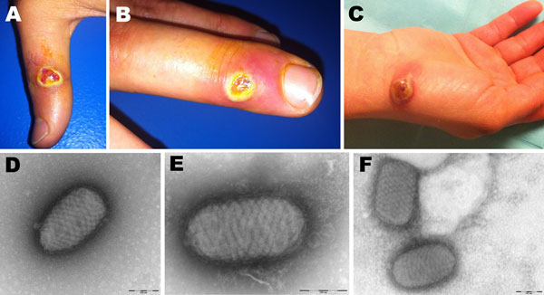
Figure 1. . Orf virus infection in 5 persons who butchered or prepared lambs as part of a religious practice for Eid al-Adha (the Muslim Feast of Sacrifice), Marseille, France, 2011. Cutaneous lesions on hands of case-patient 3 (A, B) and case-patient 5 (C) are shown. Negative-staining electron microscopy of samples from case-patient 3 (D) and case-patient 5 (E, F) show ovoid particles (≈250 nm long, 150 nm wide) with a crisscross appearance; the size and appearance of these particles are highly suggestive of parapoxvirus virions.
1These authors contributed equally to this article.
Page created: December 20, 2012
Page updated: December 20, 2012
Page reviewed: December 20, 2012
The conclusions, findings, and opinions expressed by authors contributing to this journal do not necessarily reflect the official position of the U.S. Department of Health and Human Services, the Public Health Service, the Centers for Disease Control and Prevention, or the authors' affiliated institutions. Use of trade names is for identification only and does not imply endorsement by any of the groups named above.