Volume 19, Number 12—December 2013
Dispatch
Concomitant Human Infections with 2 Cowpox Virus Strains in Related Cases, France, 2011
Corinne Ducournau, Audrey Ferrier-Rembert, Olivier Ferraris, Aurélie Joffre, Anne-Laure Favier, Olivier Flusin, Dieter Van Cauteren, Kaci Kecir, Brigitte Auburtin, Serge Védy, Maël Bessaud, and Christophe N. Peyrefitte
Figure 1
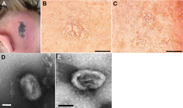
Figure 1. . Cowpox virus infection in 4 persons in France. The case-patients were infected in 2011 by virus transmitted from infected pet rats. A) Cutaneous lesion on patient 1. B) Cytopathic effects observed on Vero cell monolayers with isolate CEPAD332. Scale bar represents 500 μm. C) Cytopathic effects observed on Vero cell monolayers with isolate CEPAD335. Scale bar represents 500 μm. D) Negative-staining electron microscopy image of isolate CEPAD332. Scale bar represents 100 nm. E) Negative-staining electron microscopy image of isolate CEPAD335. Scale bar represents 100 nm.
Page created: November 19, 2013
Page updated: November 19, 2013
Page reviewed: November 19, 2013
The conclusions, findings, and opinions expressed by authors contributing to this journal do not necessarily reflect the official position of the U.S. Department of Health and Human Services, the Public Health Service, the Centers for Disease Control and Prevention, or the authors' affiliated institutions. Use of trade names is for identification only and does not imply endorsement by any of the groups named above.