Volume 19, Number 4—April 2013
Research
Circovirus in Tissues of Dogs with Vasculitis and Hemorrhage
Linlin Li, Sabrina McGraw, Kevin Zhu, Christian M. Leutenegger, Stanley L. Marks, Steven Kubiski, Patricia Gaffney, Florante N. Dela Cruz Jr, Chunlin Wang, Eric Delwart, and Patricia A. Pesavento
Figure 3
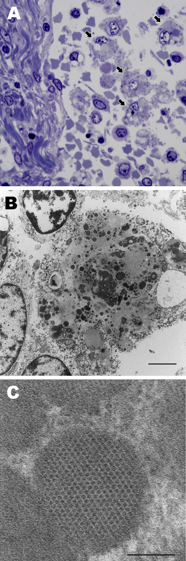
Figure 3. . . Lymph node from sentinel dog from which dog circovirus was identified. A) Toluidine blue stain shows multiple macrophages within the medullary sinus contain vacuoles and discrete, oblong to round, variably stained cytoplasmic bodies (arrows). B) A single macrophage adjacent to a lymphocyte (upper left) and partial profiles of other cells. Intracytoplasmic inclusion bodies are distributed throughout the macrophage cytoplasm, along with mitochondria and vacuoles. Scale bar indicates 2 µm. C) Intracytoplasmic inclusion bodies contain granular content and sometimes paracrystalline to herringbone arrays of 10–11 nm diameter viral-like particles. Scale bar indicates 100 nm.
Page created: March 13, 2013
Page updated: March 13, 2013
Page reviewed: March 13, 2013
The conclusions, findings, and opinions expressed by authors contributing to this journal do not necessarily reflect the official position of the U.S. Department of Health and Human Services, the Public Health Service, the Centers for Disease Control and Prevention, or the authors' affiliated institutions. Use of trade names is for identification only and does not imply endorsement by any of the groups named above.