Volume 19, Number 6—June 2013
Dispatch
BSE-associated Prion-Amyloid Cardiomyopathy in Primates
Susanne Krasemann, Giulia Mearini, Elisabeth Krämer, Katja Wagenführ, Walter Schulz-Schaeffer, Melanie Neumann, Walter Bodemer, Franz-Josef Kaup, Michael Beekes, Lucie Carrier, Adriano Aguzzi1, and Markus Glatzel1
Figure 1
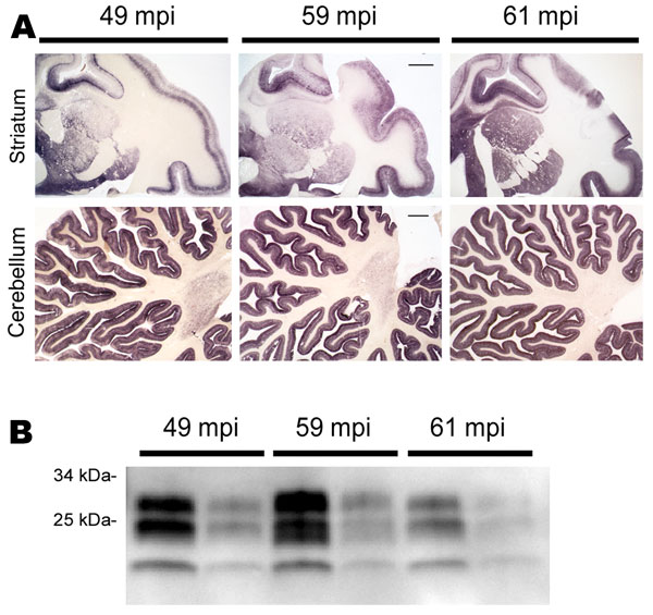
Figure 1. . PrPSc distribution and content in brain of bovine spongiform encephalopathy (BSE)–infected rhesus macaques. A) Paraffin-embedded tissue blot of striatum and cerebellum show a typical BSE-like deposition pattern of PrPSc with no differences between individual BSE-diseased monkeys at 49, 59, and 61 months postinoculation (mpi). Scale bars = 1 mm. B) Western blot analysis for PrPSc in brain of BSE-infected monkeys with incubation times of 49, 59, and 61 mpi. PrPSc-type is as expected for BSE prions, and no major differences in PrPSc load were detected. All samples were proteinase K–digested; loading amount was 0.5 and 0.1 mg fresh wet tissue for each sample
1These authors contributed equally to this article.
Page created: May 20, 2013
Page updated: May 20, 2013
Page reviewed: May 20, 2013
The conclusions, findings, and opinions expressed by authors contributing to this journal do not necessarily reflect the official position of the U.S. Department of Health and Human Services, the Public Health Service, the Centers for Disease Control and Prevention, or the authors' affiliated institutions. Use of trade names is for identification only and does not imply endorsement by any of the groups named above.