Volume 20, Number 10—October 2014
Dispatch
Human Granulocytic Anaplasmosis, South Korea, 2013
Kye-Hyung Kim1, Jongyoun Yi1, Won Sup Oh, Nak-Hyun Kim, Su Jin Choi, Pyoeng Gyun Choe, Nam-Joong Kim, Jong-Koo Lee, and Ghazi Kayali
Figure 2
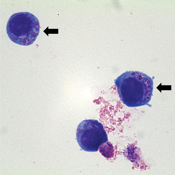
Figure 2. Light micrograph of Anaplasma phagocytophilum cultured in human promyelocytic cell line HL-60, showing A. phagocytophilum morulae as basophilic and intracytoplasmic inclusions (arrows). Wright–Giemsa stain, original magnification x1,000.
1These authors contributed equally to this article.
Page created: September 22, 2014
Page updated: September 22, 2014
Page reviewed: September 22, 2014
The conclusions, findings, and opinions expressed by authors contributing to this journal do not necessarily reflect the official position of the U.S. Department of Health and Human Services, the Public Health Service, the Centers for Disease Control and Prevention, or the authors' affiliated institutions. Use of trade names is for identification only and does not imply endorsement by any of the groups named above.