Skip directly to site content Skip directly to page options Skip directly to A-Z link Skip directly to A-Z link Skip directly to A-Z link
Volume 20, Number 11—November 2014
Dispatch

Human Herpes Simplex Virus Type 1 in Confiscated Gorilla

Kirsten V.K. GilardiComments to Author , Kristie L. Oxford, David Gardner-Roberts1, Jean-Felix Kinani, Lucy Spelman2, Peter A. Barry, Michael R. Cranfield, and Linda J. Lowenstine
Author affiliations: University of California, Davis, California, USA (K.V.K. Gilardi, K.L. Oxford, P.A. Barry, M.R. Cranfield, L.J. Lowenstine); Mountain Gorilla Veterinary Project, Inc., Davis (D. Gardner-Roberts, J.-F, Kinani, L. Spelman, M.R. Cranfield)

Main Article

Figure 1

Vesicular stomatitis in a wild-caught juvenile Grauer’s gorilla (Gorilla beringei graueri). Gross lesions, histopathologic examination, transmission electron microscopy, and molecular screening confirmed human herpesvirus type 1 (HSV-1) as the etiologic agent. A) Human HSV-1 lip lesions in a wild-caught juvenile Grauer’s gorilla. B) Section of oral mucosa adjacent to a vesiculo-ulcerative lesion exhibits epithelial cell necrosis, cytoplasmic swelling, nuclear chromatin margination (sometimes wit

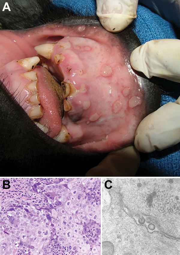
Figure 1. Vesicular stomatitis in a wild-caught juvenile Grauer’s gorilla (Gorilla beringei graueri). Gross lesions, histopathologic examination, transmission electron microscopy, and molecular screening confirmed human herpesvirus type 1 (HSV-1) as the etiologic agent. A) Human HSV-1 lip lesions in a wild-caught juvenile Grauer’s gorilla. B) Section of oral mucosa adjacent to a vesiculo-ulcerative lesion exhibits epithelial cell necrosis, cytoplasmic swelling, nuclear chromatin margination (sometimes with discrete Cowdry type A inclusions), and multinucleated syncytia typical of herpesviral cytopathic effects (hematoxylin and eosin stain). Original magnification ×200. C) Electron micrograph of the same lesion demonstrates intranuclear, unenveloped virions ≈100 nm in diameter that are budding through the nuclear membrane to form enveloped virions ≈140 nm in diameter; morphologic features of both are compatible with a herpesvirus. Original magnification ×60,000.

Main Article

1Current affiliation: ARMAC Veterinary Group, Biggar, South Lanarkshire, Scotland, UK.

2Current affiliation: Rhode Island School of Design, Providence, Rhode Island, USA.

Page created: October 17, 2014
Page updated: October 17, 2014
Page reviewed: October 17, 2014
The conclusions, findings, and opinions expressed by authors contributing to this journal do not necessarily reflect the official position of the U.S. Department of Health and Human Services, the Public Health Service, the Centers for Disease Control and Prevention, or the authors' affiliated institutions. Use of trade names is for identification only and does not imply endorsement by any of the groups named above.
file_external