Volume 20, Number 2—February 2014
Synopsis
Anncaliia algerae Microsporidial Myositis
Matthew R. Watts , Renee C.F. Chan, Elaine Y.L. Cheong, Susan Brammah, Kate R. Clezy, Chiwai Tong, Deborah Marriott, Cameron E. Webb, Bobby Chacko, Vivienne Tobias, Alexander C. Outhred, Andrew S. Field, Michael V. Prowse, James V. Bertouch, Damien Stark, and Stephen W. Reddel
Figure 2
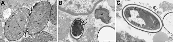
Figure 2. Electron micrographs of muscle biopsy tissue from a 66-year-old man (case-patient B) (A) and a 67-year-old man (case-patient A) (B,C) showing Anncaliia algerae. A) Early proliferative stage meronts with diplokaryotic nuclei (n) and vesiculotubular appendages (arrows) attached to the plasmalemmaScale bar indicates 1 μmB) Degenerate crenated sporoblast (sb) and a mature spore with visible coils of the polar tubule (arrows)Scale bar indicates 1 μmC) Mature spore with 9 polar tubule coils in a single row, pale endospore, and a dense exosporeScale bar indicates 500 nm.
Page created: January 17, 2014
Page updated: January 17, 2014
Page reviewed: January 17, 2014
The conclusions, findings, and opinions expressed by authors contributing to this journal do not necessarily reflect the official position of the U.S. Department of Health and Human Services, the Public Health Service, the Centers for Disease Control and Prevention, or the authors' affiliated institutions. Use of trade names is for identification only and does not imply endorsement by any of the groups named above.