Pneumonic Plague Outbreak, Northern Madagascar, 2011
Vincent Richard, Julia M. Riehm

, Perlinot Herindrainy, Rahelinirina Soanandrasana, Maherisoa Ratsitoharina, Fanjasoa Rakotomanana, Samuel Andrianalimanana, Holger C. Scholz, and Minoarisoa Rajerison
Author affiliations: Institut Pasteur de Dakar, Dakar, Senegal (V. Richard); Bundeswehr Institute of Microbiology, Munich, Germany (J.M. Riehm, H.C. Scholz); German Center for Infection Research, Munich (J.M. Riehm, H.C. Scholz); Institut Pasteur de Madagascar, Anatananarivo, Madagascar (P. Herindrainy, R. Soanandrasana, M. Ratsitoharina, F. Rakotomanana, M. Rajerison); Ministry of Public Health, Anatananarivo (S. Andrianalimanana)
Main Article
Figure 2
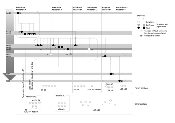
Figure 2. Infection pattern during pneumonic plague outbreak, northern Madagascar, 2011. The outbreak spread to other neighboring villages during January 14–February 9. Twenty persons in 6 households (A–F) in 5 villages had symptoms of pneumonic plague. The outbreak population was divided into 3 groups (group 1: case-patients 1–5; group 2: case-patients 6–17; and group 3: case-patients 18–20). Patients received treatment by January 28. Because of geographic distance, none of the patients in group 3 received treatment. Contacts were divided into family contacts (c1–c16) who lived in an affected household and other contacts (c17–c41) who interacted with infected patients or patients who died. All contacts, except c10 and c25, received antimicrobial drug prophylaxis. Two contacts (c14 and c25) were seropositive (single serum sample); all other contacts remained seronegative.
Main Article
Page created: December 17, 2014
Page updated: December 17, 2014
Page reviewed: December 17, 2014
The conclusions, findings, and opinions expressed by authors contributing to this journal do not necessarily reflect the official position of the U.S. Department of Health and Human Services, the Public Health Service, the Centers for Disease Control and Prevention, or the authors' affiliated institutions. Use of trade names is for identification only and does not imply endorsement by any of the groups named above.