Volume 21, Number 4—April 2015
Dispatch
Pathogenicity of 2 Porcine Deltacoronavirus Strains in Gnotobiotic Pigs
Figure 2
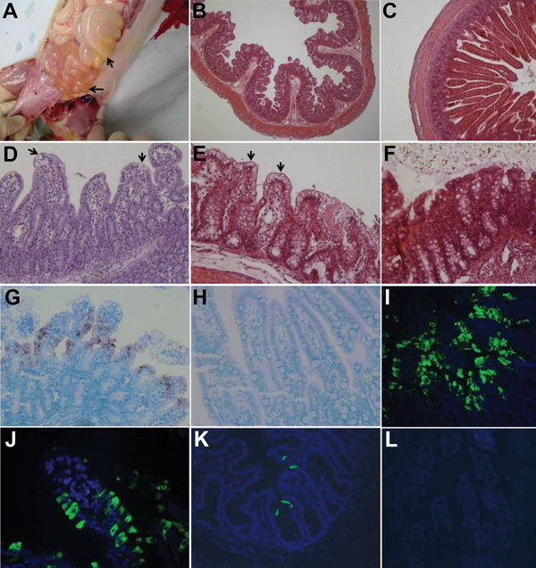
Figure 2. Intestinal changes in gnotobiotic pigs inoculated with porcine deltacoronavirus (PDCoV) strains OH-FD22 (panels A, B, E, G, I, and J) and OH-FD100 (panels D and K). A) Intestine of pig 3 at hour postinoculation (hpi) 72 (48–51 h after onset of clinical signs), showing thin and transparent intestinal walls (duodenum to colon) and accumulation of large amounts of yellow fluid in the intestinal lumen (arrows). B) Jejunum of pig 3 at hpi 72 (48–51 h after onset of clinical signs), showing acute diffuse, severe atrophic jejunitis (original magnification ×40). C) Jejunum of noninoculated pig 7, showing normal villous epithelium (original magnification ×80). D) Jejunum of pig 4 at hpi 96 (72–74 h after onset of clinical signs), showing acute diffuse, severe atrophic jejunitis with mild cytoplasmic vacuolation at the tips of villi (arrows) (original magnification ×200). E) Colon of pig 3 at hpi 72 (48–51 h after onset of clinical signs), showing mild cytoplasmic vacuolation of superficial epithelial cells (arrows) (original magnification ×200). F) Colon of noninoculated pig 7, showing normal colonic epithelium (original magnification ×200). G) Jejunum of pig 3 at hpi 72 (48–51 h after onset of clinical signs), showing that epithelial cells lining the atrophied villi are positive for PDCoV RNA (original magnification ×200). H) Jejunum of noninoculated pig 6, showing absence of PDCoV RNA-positive cells and background staining (original magnification ×200). I) Jejunum of pig 3 at hpi 72 (48–51 h after onset of clinical signs), showing large numbers of PDCoV antigen–positive cells in the epithelium of atrophied villi (original magnification ×200). J) Jejunum of pig 3 at hpi 72 (48–51 h after onset of clinical signs), showing localization of PDCoV antigens in cytoplasm of columnar epithelial cells (original magnification ×400). K) Cecum of pig 4 at hpi 96 (72–74 h after onset of clinical signs), showing a few PDCoV antigen–positive cells in the epithelium (original magnification ×200. L) Jejunum of noninoculated pig 6, showing absence of immunofluorescence-stained cells and background staining (original magnification ×200). Nuclei were stained with blue-fluorescent 4′, 6-diamidino-2-phenylindole dihydrochloride. Hematoxylin and eosin staining in panels B–F; in situ hybridization staining in panels G and H; immunofluorescence staining in panels I–L.