Recent US Case of Variant Creutzfeldt-Jakob Disease—Global Implications
Atul Maheshwari

, Michael Fischer, Pierluigi Gambetti, Alicia Parker, Aarthi Ram, Claudio Soto, Luis Concha-Marambio, Yvonne Cohen, Ermias D. Belay, Ryan A. Maddox, Simon Mead, Clay Goodman, Joseph S. Kass, Lawrence B. Schonberger, and Haitham M. Hussein
Author affiliations: Baylor College of Medicine, Houston, Texas, USA (A. Maheshwari, A. Parker, A. Ram, C. Goodman, J.S. Kass); Harris Health System, Houston (A. Maheshwari, A. Parker, A. Ram, J.S. Kass); Texas Department of State Health Services, Austin, Texas, USA (M. Fischer); Case Western Reserve University School of Medicine, Cleveland, Ohio, USA (P. Gambetti, Y. Cohen); University of Texas Medical School at Houston, Houston (C. Soto, L. Concha-Marambio); Universidad de los Andes, Santiago, Chile (L. Concha-Marambio); Centers for Disease Control and Prevention, Atlanta, Georgia, USA (E.D. Belay, R.A. Maddox, L.B. Schonberger); University College London Institute of Neurology, London, UK (S. Mead); HealthPartners Clinics & Services, St. Paul, Minnesota, USA (H.M. Hussein)
Main Article
Figure 3
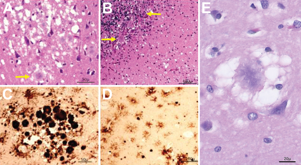
Figure 3. Results of histopathologic and immunohistochemical analyses for a US patient with variant Creutzfeldt-Jakob disease. A) Hematoxylin and eosin staining shows many typical florid plaques (A, arrow) occasionally forming clusters; large vacuole spongiform change is also present (original magnification ×10). B) Plaques often not of the florid type along with spongiform change are present in cerebellum (arrows; original magnification ×20). C, D) Prion protein immunostaining confirms the presence of PrP in the plaques, which intensely immunostained (C; original magnification ×10), and highlights small round cells surrounded by short processes (D, antibody 3F4; original magnification ×40). E) Under high magnification, a prion plaque with spongiform change is seen in the left frontal cortex (original magnification ×400).
Main Article
Page created: April 19, 2015
Page updated: April 19, 2015
Page reviewed: April 19, 2015
The conclusions, findings, and opinions expressed by authors contributing to this journal do not necessarily reflect the official position of the U.S. Department of Health and Human Services, the Public Health Service, the Centers for Disease Control and Prevention, or the authors' affiliated institutions. Use of trade names is for identification only and does not imply endorsement by any of the groups named above.