Volume 22, Number 1—January 2016
Dispatch
Porcine Epidemic Diarrhea Virus and Discovery of a Recombinant Swine Enteric Coronavirus, Italy
Abstract
Porcine epidemic diarrhea virus (PEDV) has been detected sporadically in Italy since the 1990s. We report the phylogenetic relationship of swine enteric coronaviruses collected in Italy during 2007–2014 and identify a drastic shift in PEDV strain variability and a new swine enteric coronavirus generated by recombination of transmissible gastroenteritis virus and PEDV.
Porcine epidemic diarrhea virus (PEDV) and Transmissible gastroenteritis virus (TGEV) (family Coronaviridae, genus Alphacoronavirus) are enveloped viruses that contain a single-stranded, positive-sense RNA genome of ≈28 kb. Infection with these viruses causes watery diarrhea, dehydration, and a high mortality rate among suckling pigs. Coronaviruses (CoVs) are prone to genetic evolution through accumulation of point mutations and homologous recombination among members of the same genus (1). Porcine respiratory coronavirus (PRCV), a mutant of TGEV, appeared in pigs in the 1980s (2). The spread of PRCV, which conserved most of the antigenic sites and causes cross-protection against TGEV (3), led to the gradual disappearance of TGEV. Newly emerging CoVs pose a potential threat to human and animal health because multiple human CoV infections have been derived from animal hosts. Emerging swine coronaviruses are of great concern to swine health because of the potential increase in viral pathogenesis.
In 1978, PEDV was first identified in Europe; subsequent reports occurred in many countries in Asia, including China, Japan, Korea, and Thailand. In 2010–2012, genetically different PEDV strains emerged, causing severe outbreaks in China (4). PEDV spread to the United States, Canada, and Mexico in 2013–2014, and genetically related strains were detected in South Korea and Taiwan (5–7). The PEDV outbreak caused large global economic losses to the swine industry. In Europe, a severe PEDV epidemic occurred in Italy during 2005–2006 (8), and in 2014–2015, PEDV was detected in Germany, France, and Belgium. These strains have a high nucleotide identity to PEDV strains that contain distinct insertions and deletions (INDELs) in the S gene (S-INDELs) from the United States (9–11). We report the detection and genetic characterization of swine enteric CoVs circulating in Italy during 2007–2014. We also report a recombinant TGEV and PEDV strain (identified as the species Swine enteric coronavirus [SeCoV]) circulating from June 2009 through 2012. Finally, we describe the phylogenetic relationship of the 2014 PEDV S-INDELs to the recent PEDV strains circulating in Europe.
During 2007–2014, we collected 27 fecal and 24 intestinal samples from pigs with suspected PEDV or TGEV infections; the pigs came from swine farms in northern Italy (Po Valley), which contains the regions of Piemonte, Lombardia, Emilia Romagna, and Veneto (Technical Appendix Figure 1). The Po Valley contains 70% of Italy’s swine. Clinical signs included watery diarrhea in sows and a death rate in piglets of 5%–10%, lower than is typical with PEDV or TGEV infections. Samples were submitted for testing by electron microscopy, PEDV ELISA, viral isolation, pan-CoV reverse transcription PCR (RT-PCR), and RT-PCR for PRCV and TGEV; selected positive pan-CoV samples were sequenced (12–14) (Technical Appendix).
Results of electron microscopy showed that 25 (49%) of the 51 samples contained CoV-like particles, but all samples were negative for viral isolation. Although only 38 samples (74%) were positive by pan-CoV RT-PCR, 47 (92%) were positive by the PEDV ELISA (Table 1) (12,13). Of the 38 pan-CoV–positive samples, 18 were selected for partial RNA-dependent RNA polymerase (RdRp), spike (S1) (14), and membrane (M) sequencing (Table 1). All samples were negative for PRCV and TGEV by RT-PCR, ruling out co-infection with PEDV and TGEV or PRCV (15).
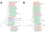
On the basis of the partial sequences from RdRp and the S1 and M genes, the strains from Italy clustered into 3 temporally divided groups, suggesting 3 independent virus entries. Cluster I represents strains circulating from 2007 through mid-2009; cluster II represents strains circulating from mid-2009 through 2012; and cluster III represents strains circulating since 2014 (Technical Appendix Figure 2, panels A–C). Cluster I was identified in Emilia Romagna (n = 1), Lombardia (n = 5), and Veneto (n = 1). Cluster II was identified in Emilia Romagna (n = 1) and Lombardia (n = 8). Cluster III was identified in Emilia Romagna at 2 swine farms. To help explain the temporal clustering, a single S1 gene segment was sequenced from clusters I and II (PEDV/Italy/7239/2009 and SeCoV/Italy/213306/2009, respectively). Because of the recent outbreak of PEDV in Europe, the 2 positive samples from cluster III (PEDV/Italy/178509/2014 and PEDV/Italy/200885/2014) were sequenced (Figure 1, panel A).
One strain from each cluster was selected for whole genome sequencing (Technical Appendix). Unfortunately, the whole genome was obtained from only clusters I and II (PEDV/Italy/7239/2009 and SeCoV/Italy/213306/2009, respectively; Figure 1, panel B). Recombination analysis was conducted on the 2 whole genomes and was not detected in PEDV/Italy/7239/2009. Recombination was detected in SeCoV/Italy/213306/2009 at position 20636 and 24867 of PEDV CV777 and at position 20366 and 24996 of TGEV H16 (Figure 2), suggesting the occurrence of a recombination event between a PEDV and a TGEV. The complete S gene of SeCoV/Italy/213306/2009 shared 92% and 90% nt identity with the prototype European strain PEDV CV777 and the original highly virulent North American strain Colorado 2013, respectively, and the remaining genome shared a 97% nt identity with the virulent strains TGEV H16 and TGEV Miller M6. Whole-genome analysis of PEDV/Italy/7239/2009 showed that it grouped with the global PEDV strains (6) and shared ≈97% nt identity with PEDV strains CV777, DR13 virulent, and North American S-INDEL strain OH851 (Table 2).
During 2007–2014, most (92%) samples collected from the Po Valley in Italy were positive for PEDV by ELISA; only 72% were positive by pan-CoV PCR. However, because we were investigating the presence of PEDV or TGEV in samples with clinical signs of diarrhea, the high occurrence of PEDV may not reflect the actual prevalence of PEDV in Italy. The increased percentage of PEDV found in samples tested by ELISA, compared with the proportion found by PCR, may be explained by the number of ambiguous bases in the pan-CoV primers; the ambiguous bases severely reduce the efficiency of the reaction. The swine enteric CoV strains from Italy in our study, including the recombinant strain, were reported in pigs with mild clinical signs, indicating that PEDV and SeCoV have been circulating in Italy and likely throughout Europe for multiple years but were underestimated as a mild form of diarrhea.
To understand the evolution of PEDV in Italy, the partial RdRp, S, and M genes were sequenced from 18 samples and grouped in 3 different temporal clusters. Cluster I (2007–mid 2009) resembles the oldest PEDV strains; cluster II resembles a new TGEV and PEDV recombinant variant; and cluster III, identified from 2 pig farms in northern Italy in 2014, resembles the PEDV S-INDEL strains identified in Germany, France, Belgium, and the United States. The >99.3% nt identity of the S1 gene within cluster III and in previously identified strains could suggest the spread of the S-INDEL strain into Europe. However, directionality of spread cannot be determined because of a lack of global and temporal PEDV sequences.
Although our findings could indicate 3 introductions of PEDV in Italy, the results more likely suggest the high ability of natural recombination among CoVs and the continued emergence of novel CoVs with distinct pathogenic properties. Further investigation is needed to determine the ancestor of the SeCoV strain or to verify whether the recombinant virus was introduced in Italy. Recombinant SeCoV was probably generated in a country in which both PEDV and TGEV are endemic, but because the presence of these viruses in Europe is unclear and SeCoV has not been previously described, it is difficult to determine the parental strains and geographic spread of SeCoV. Future studies are required to describe the pathogenesis of SeCoV and its prevalence in other countries.
Dr. Boniotti is a scientist at the Istituto Zooprofilattico Sperimentale della Lombardia e Emilia Romagna; her primary research interests include molecular diagnosis and epidemiology of viral and bacterial infectious diseases. Dr. Papetti is a research scientist at the Istituto Zooprofilattico Sperimentale della Lombardia e Emilia Romagna; her major research interests are molecular diagnostic development and phylogenetic analysis of infectious diseases.
Acknowledgment
This work was funded by the Italian Ministry of Health (project PRC2010010- E87G11000130001).
References
- Enjuanes L, Brian D, Cavanagh D, Holmes K, Lai MMC, Laude H, Coronaviridae. In: van Regenmortel MHV, Fauquet CM, Bishop DHL, Carstens EB, Estes MK, Lemon SM, et al., editors. Virus taxonomy. Classification and nomenclature of viruses. New York: Academic Press; 2000. p. 835–9.
- Pensaert M, Callebaut P, Vergote J. Isolation of a porcine respiratory, non-enteric coronavirus related to transmissible gastroenteritis. Vet Q. 1986;8:257–61. DOIPubMedGoogle Scholar
- Wesley RD, Woods RD. Immunization of pregnant gilts with PRCV induces lactogenic immunity for protection of nursing piglets from challenge with TGEV. Vet Microbiol. 1993;38:31–40. DOIPubMedGoogle Scholar
- Huang YW, Dickerman AW, Py ñeyro P, Li L, Fang L, Kiehne R, Origin, evolution, and genotyping of emergent porcine epidemic diarrhea virus strains in the United States. mBiol. 2013;4:00737-13. DOIPubMedGoogle Scholar
- Stevenson GW, Hoang H, Schwartz KJ, Burrough ER, Sun D, Madson D, Emergence of porcine epidemic diarrhea virus in the United States: clinical signs, lesions, and viral genomic sequences. J Vet Diagn Invest. 2013;25:649–54. DOIPubMedGoogle Scholar
- Vlasova AN, Marthaler D, Wang Q, Culhane MR, Rossow KD, Rovira A, Distinct characteristics and complex evolution of PEDV strains, North America, May 2013–February 2014. Emerg Infect Dis. 2014;20:1620–8. DOIPubMedGoogle Scholar
- Sun M, Ma J, Wang Y, Wang M, Song W, Zhang W, Genomic and epidemiological characteristics provide new insights into the phylogeographical and spatiotemporal spread of porcine epidemic diarrhea virus in Asia. J Clin Microbiol. 2015;53:1484–92. DOIPubMedGoogle Scholar
- Martelli P, Lavazza A, Nigrelli AD, Merialdi G, Alborali LG, Pensaert MB. Epidemic of diarrhoea caused by porcine epidemic diarrhoea virus in Italy. Vet Rec. 2008;162:307–10. DOIPubMedGoogle Scholar
- Hanke D, Jenckel M, Petrov A, Ritzmann M, Stadler J, Akimkin V, Comparison of porcine epidemic diarrhea viruses from Germany and the United States, 2014. Emerg Infect Dis. 2015;21:493–6. DOIPubMedGoogle Scholar
- Grasland B, Bigault L, Bernard C, Quenault H, Toulouse O, Fablet C, Complete genome sequence of a porcine epidemic diarrhea S gene indel strain isolated in France in December 2014. Genome Announc. 2015;3:e00535-15. DOIPubMedGoogle Scholar
- Theuns S, Conceição-Neto N, Christiaens I, Zeller M, Desmarets LMB, Roukaerts IDM, Complete genome sequence of a porcine epidemic diarrhea virus from a novel outbreak in Belgium, January 2015. Genome Announc. 2015;3:e00506-15. DOIPubMedGoogle Scholar
- Sozzi E, Luppi A, Lelli D, Moreno Martin A, Canelli E, Brocchi E, Comparison of enzyme-linked immunosorbent assay and RT-PCR or the detection of porcine epidemic diarrhoea virus. Res Vet Sci. 2010;88:166–8. DOIPubMedGoogle Scholar
- Lelli D, Papetti A, Sabelli C, Rosti E, Moreno A, Boniotti MB. Detection of coronaviruses in various bat species in Italy. Viruses. 2013;5:2679–89. DOIPubMedGoogle Scholar
- Kim SY, Song DS, Park BK. Differential detection of transmissible gastroenteritis virus and porcine epidemic diarrhea virus by duplex RT-PCR. J Vet Diagn Invest. 2001;13:516–20. DOIPubMedGoogle Scholar
- Kim L, Chang KO, Sestak K, Parwani A, Saif LJ. Development of a reverse transcription–nested polymerase chain reaction assay for differential diagnosis of transmissible gastroenteritis virus and porcine respiratory coronavirus from feces and nasal swabs of infected pigs. J Vet Diagn Invest. 2000;12:385–8. DOIPubMedGoogle Scholar
Figures
Tables
Cite This Article1These first authors contributed equally to this article.
2Deceased.
Table of Contents – Volume 22, Number 1—January 2016
| EID Search Options |
|---|
|
|
|
|
|
|


Please use the form below to submit correspondence to the authors or contact them at the following address:
M. Beatrice Boniotti, Research and Development Laboratory, Istituto Zooprofilattico Sperimentale della Lombardia e Emilia Romagna, via Bianchi 9, 25124 Brescia (BS), Italy
Top