Skip directly to site content Skip directly to page options Skip directly to A-Z link Skip directly to A-Z link Skip directly to A-Z link
Volume 22, Number 4—April 2016
Letter

Sustained Elevated Cytokine Levels during Recovery Phase of Mayaro Virus Infection

Dennis TappeComments to Author , José Vicente Pérez-Girón, Gudrun Just-Nübling, Gernot Schuster, Sergio Gómez-Medina, Stephan Günther, César Muñoz-Fontela, and Jonas Schmidt-Chanasit
Author affiliations: Bernhard Nocht Institute for Tropical Medicine, Hamburg, Germany (D. Tappe, S. Günther, C. Muñoz-Fontela, J. Schmidt-Chanasit); German Centre for Infection Research, Hamburg (D. Tappe, S. Günther, C. Muñoz-Fontela, J. Schmidt-Chanasit); Leibniz Institute for Experimental Virology, Hamburg (J. Vicente Pérez-Girón, S. Gómez-Medina, C. Muñoz-Fontela); J.W. Goethe University Center for Infectious and Tropical Diseases, Frankfurt, Germany (G. Just-Nübling); Praxis Schuster, Reinhardshagen, Germany (G. Schuster)

Main Article

Figure

Changes in cytokine and growth factor levels in the acute and recovery phase of Mayaro fever. Box-and-whisker plots show median, upper and lower quartile, minimum, and maximum values. A) During the prolonged recovery phase, serum levels of interleukin (IL) 5–10, IL-13, and IL-17 were significantly elevated compared with levels for healthy controls. IL-10 levels were also significantly increased during the acute phase, as were IL-12p70 levels. B) Significantly increased serum concentrations of in

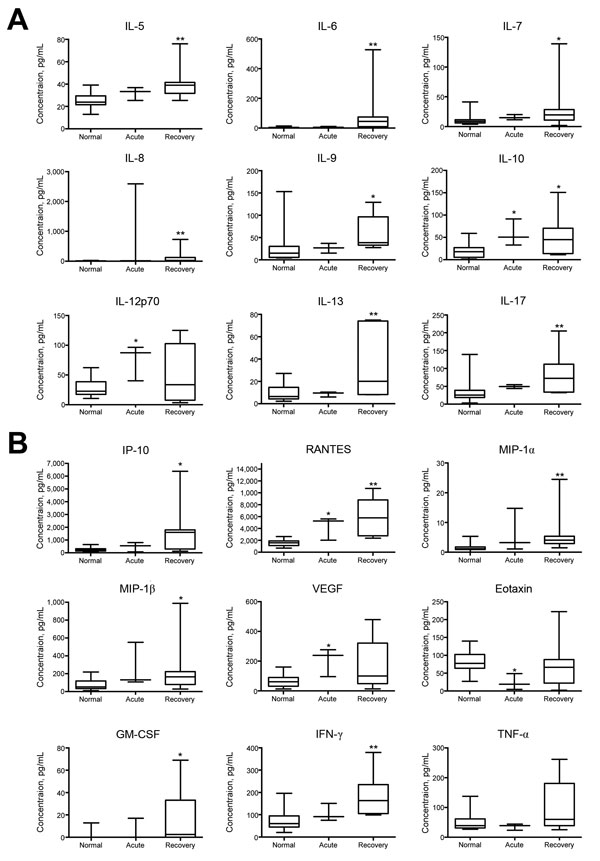
Figure. Changes in cytokine and growth factor levels in the acute and recovery phase of Mayaro fever. Box-and-whisker plots show median, upper and lower quartile, minimum, and maximum values. A) During the prolonged recovery phase, serum levels of interleukin (IL) 5–10, IL-13, and IL-17 were significantly elevated compared with levels for healthy controls. IL-10 levels were also significantly increased during the acute phase, as were IL-12p70 levels. B) Significantly increased serum concentrations of interferon-γ (IFN-γ)–induced protein 10 (IP-10), regulated on activation, normal T cell expressed and secreted (RANTES), macrophage inflammatory proteins (MIP)–1α and –1β, granulocyte-macrophage colony-stimulating factor (GM-CSF), and IFN-γ were detected in the prolonged recovery phase; significant elevations were also seen in the acute phase for RANTES and vascular endothelial growth factor (VEGF). Tumor necrosis factor α (TNF-α) concentrations showed a nonsignificant median decrease during the acute phase, whereas eotaxin levels were significantly decreased at that time. *p<0.05 and **p<0.01, versus values for healthy controls (Kruskal-Wallis test).

Main Article

Page created: March 16, 2016
Page updated: March 16, 2016
Page reviewed: March 16, 2016
The conclusions, findings, and opinions expressed by authors contributing to this journal do not necessarily reflect the official position of the U.S. Department of Health and Human Services, the Public Health Service, the Centers for Disease Control and Prevention, or the authors' affiliated institutions. Use of trade names is for identification only and does not imply endorsement by any of the groups named above.
file_external