Skip directly to site content Skip directly to page options Skip directly to A-Z link Skip directly to A-Z link Skip directly to A-Z link
Volume 22, Number 6—June 2016
Research

Infection, Replication, and Transmission of Middle East Respiratory Syndrome Coronavirus in Alpacas

Danielle R. Adney, Helle Bielefeldt-Ohmann, Airn E. Hartwig, and Richard A. BowenComments to Author 
Author affiliations: Colorado State University, Fort Collins, Colorado, USA (D.R. Adney, A.E. Hartwig, R.A. Bowen); University of Queensland, Gatton, Queensland, Australia (H. Bielefeldt-Ohmann)

Main Article

Figure 4

Signs of mild upper respiratory inflammation, encephalitis, and virus antigen detection in respiratory epithelium of alpacas experimentally infected with Middle East respiratory syndrome coronavirus. A) Turbinate from alpaca A8 showing normal respiratory epithelium on the right with goblet cells (blue cells). Epithelium on the left has undergone squamous metaplasia (arrows) and is focally eroded with mild subepithelial inflammation (original magnification ×100). B) Virus antigen in apparently in

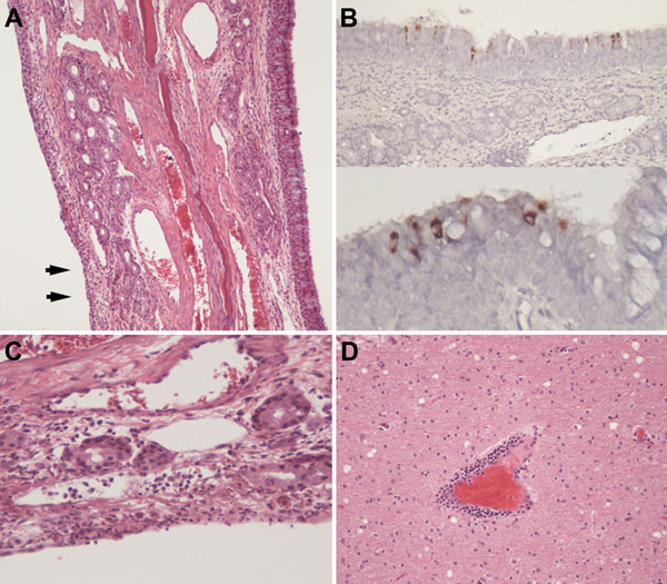
Figure 4. Signs of mild upper respiratory inflammation, encephalitis, and virus antigen detection in respiratory epithelium of alpacas experimentally infected with Middle East respiratory syndrome coronavirus. A) Turbinate from alpaca A8 showing normal respiratory epithelium on the right with goblet cells (blue cells). Epithelium on the left has undergone squamous metaplasia (arrows) and is focally eroded with mild subepithelial inflammation (original magnification ×100). B) Virus antigen in apparently intact respiratory epithelium of alpaca A8 detected by immunohistochemical analysis and lack of subepithelial inflammation (original magnification ×200 [top] and ×400 [bottom]). C) Erosion in turbinate epithelium from alpaca A8 showing leukocytosis in underlying blood vessels (original magnification ×400). D) Perivascular infiltration of lymphocytes and monocytes in the brain of alpaca A9 (original magnification ×200). Hematoxylin and eosin stain in panels A, B, and D; immunohistochemical stain in panel C.

Main Article

Page created: May 16, 2016
Page updated: May 16, 2016
Page reviewed: May 16, 2016
The conclusions, findings, and opinions expressed by authors contributing to this journal do not necessarily reflect the official position of the U.S. Department of Health and Human Services, the Public Health Service, the Centers for Disease Control and Prevention, or the authors' affiliated institutions. Use of trade names is for identification only and does not imply endorsement by any of the groups named above.
file_external