Volume 23, Number 2—February 2017
Perspective
Delivering on Antimicrobial Resistance Agenda Not Possible without Improving Fungal Diagnostic Capabilities
David W. Denning , David S. Perlin, Eavan G. Muldoon, Arnaldo Lopes Colombo, Arunaloke Chakrabarti, Malcolm D. Richardson, and Tania C. Sorrell
Figure 3
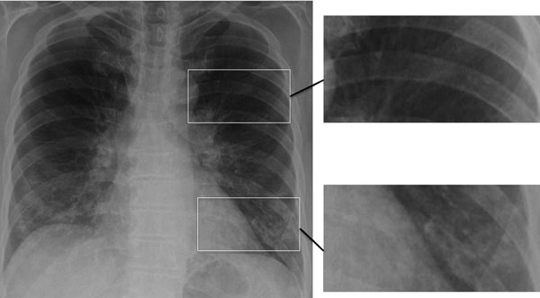
Figure 3. Chest radiograph showing early, subtle Pneumocystis pneumonia–associated abnormalities in both lower lungs of a patient newly diagnosed with AIDS; this diagnosis was unsuspected in the patient, a 63-year-old married man. Magnified images on right show normal lung (top image) and infiltrates adjacent to and behind the heart and overlain by rib (bottom image). Similar differences between the upper and lower lobes are seen in the radiograph on the left. Image used with permission of David Denning (©2016, all rights reserved).
Page created: January 17, 2017
Page updated: January 17, 2017
Page reviewed: January 17, 2017
The conclusions, findings, and opinions expressed by authors contributing to this journal do not necessarily reflect the official position of the U.S. Department of Health and Human Services, the Public Health Service, the Centers for Disease Control and Prevention, or the authors' affiliated institutions. Use of trade names is for identification only and does not imply endorsement by any of the groups named above.