Skip directly to site content Skip directly to page options Skip directly to A-Z link Skip directly to A-Z link Skip directly to A-Z link
Volume 23, Number 2—February 2017
Dispatch

Biofilm-Forming Capability of Highly Virulent, Multidrug-Resistant Candida auris

Leighann Sherry, Gordon Ramage, Ryan Kean, Andrew Borman, Elizabeth M. Johnson, Malcolm D. Richardson, and Riina Rautemaa-RichardsonComments to Author 
Author affiliations: University of Glasgow, Glasgow, Scotland, UK (L. Sherry, G. Ramage, R. Kean); Public Health England, Bristol, UK (A. Borman, E.M. Johnson); The University of Manchester, Manchester, UK (M.D. Richardson, R. Rautemaa-Richardson); University Hospital of South Manchester, Manchester (M.D. Richardson, R. Rautemaa-Richardson)

Main Article

Figure 1

Biofilm formation on Candida auris, C. albicans, and C. glabrata yeast strains. A) Biomass quantities were determined spectrophotometrically for 4 strains of C. auris and 1 each of C. albicans and C. glabrata. Isolates were standardized to 106 cells/mL in RPMI-1640 and grown in flat-bottomed 96-well microtiter plates for 24 h at 37°C. Biofilms were then washed, stained with crystal violet solution, and quantified. Data represent the mean ± SD of experiments performed on 3 separate occasions, usi

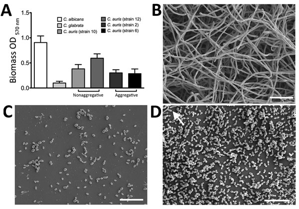
Figure 1. Biofilm formation on Candida auris, C. albicans, and C. glabrata yeast strains. A) Biomass quantities were determined spectrophotometrically for 4 strains of C. auris and 1 each of C. albicans and C. glabrata. Isolates were standardized to 106 cells/mL in RPMI-1640 and grown in flat-bottomed 96-well microtiter plates for 24 h at 37°C. Biofilms were then washed, stained with crystal violet solution, and quantified. Data represent the mean ± SD of experiments performed on 3 separate occasions, using 8 replicates for each strain. Results show that C. auris can form heterogeneous intermediate biofilms. B–D) C. albicans (B), C. glabrata (C) and C. auris (D) were also grown on Thermanox coverslips (Thermo Fisher Scientific, Paisley, UK) for 24 h at 37°C. Biofilms were then processed and viewed on a JEOL (Tokyo, Japan) JSM-6400 scanning electron microscope; images were assembled using Photoshop software (http://www.photoshop.com/products). Arrow indicates pseudohyphae in C. auris biofilm (D). Scale bars represent 20 μm (original magnification ×1,000). OD, optical density.

Main Article

Page created: January 17, 2017
Page updated: January 17, 2017
Page reviewed: January 17, 2017
The conclusions, findings, and opinions expressed by authors contributing to this journal do not necessarily reflect the official position of the U.S. Department of Health and Human Services, the Public Health Service, the Centers for Disease Control and Prevention, or the authors' affiliated institutions. Use of trade names is for identification only and does not imply endorsement by any of the groups named above.
file_external