Volume 23, Number 2—February 2017
Dispatch
Oral Transmission of L-Type Bovine Spongiform Encephalopathy Agent among Cattle
Figure 1
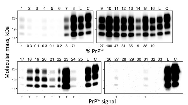
Figure 1. Western blot analysis of proteinase K–resistant disease-associated prion protein (PrPSc) in tissue samples obtained from a cow at 88 months after oral inoculation with brain homogenate of L-type bovine spongiform encephalopathy (BSE) agent. The tissues tested are shown by lane: 1, olfactory bulb; 2, frontal cortex; 3, piriform cortex; 4, parietal cortex; 5, occipital cortex; 6, hippocampus; 7, putamen; 8, thalamus; 9, hypothalamus; 10, midbrain (superior colliculus); 11, obex; 12, cervical enlargement (C7) of spinal cord; 13, lumbar enlargement (L5) of spinal cord; 14, cerebellar cortex; 15, cerebellar white matter; 16, retina; 17, neurohypophysis; 18, trigeminal ganglion; 19, dorsal root ganglion (L5); 20, cervical cranial ganglion; 21, stellate ganglion; 22, celiac and mesenteric ganglion complex; 23, optic nerve; 24, cauda equina; 25, facial nerve; 26, hypoglossal nerve; 27, cervical vagus nerve; 28, sympathetic chain; 29, brachial nerve plexus; 30, sciatic nerve; 31, adrenal gland (medulla); 32, ileum; 33, colon. Lanes 1–16 and lanes 17–33 were loaded with 0.5 mg and 100 mg tissue equivalent, respectively. As controls, lanes L and C were also loaded with 0.5 mg of L-BSE and 0.17 mg of C-BSE cattle brain equivalent, respectively. The relative percentages of PrPSc (below each lane, upper panel) are normalized against midbrain. The PrPSc signals in the extracerebral tissues (below each lane, lower panel) are indicated as positive (+) or negative (–).