Skip directly to site content Skip directly to page options Skip directly to A-Z link Skip directly to A-Z link Skip directly to A-Z link
Volume 23, Number 2—February 2017
CME ACTIVITY - Research

Multidrug-Resistant Candida haemulonii and C. auris, Tel Aviv, Israel

Ronen Ben-AmiComments to Author , Judith Berman, Ana Novikov, Edna Bash, Yael Shachor-Meyouhas, Shiri Zakin, Yasmin Maor, Jalal Tarabia, Vered Schechner, Amos Adler, and Talya Finn
Author affiliations: Tel Aviv University, Tel Aviv, Israel (R. Ben-Ami, J. Berman, S. Zakin, Y. Maor, V. Schechner, A. Adler); Tel Aviv Sourasky Medical Center, Tel Aviv (R. Ben-Ami, A. Novikov, E. Bash, J. Tarabia, V. Schechner, A. Adler, T. Finn); Ruth Rappaport Children's Hospital, Haifa, Israel (Y. Shachor-Meyouhas); Wolfson Medical Center, Holon, Israel (Y. Maor).

Main Article

Figure 4

Differing virulence of Candida auris and C. haemulonii assessed in a mouse model of hematogenous disseminated candidiasis. Virulence was assessed in immunosuppressed BALB/c mice after intravenous injection of yeast cell suspension. A) Survival curves showing significantly shorter survival of mice infected with C. albicans than C. auris and no death among mice infected with C. haemulonii. B) Kidney fungal load (CFU per gram of tissue) shown to be significantly higher in mice infected with C. albi

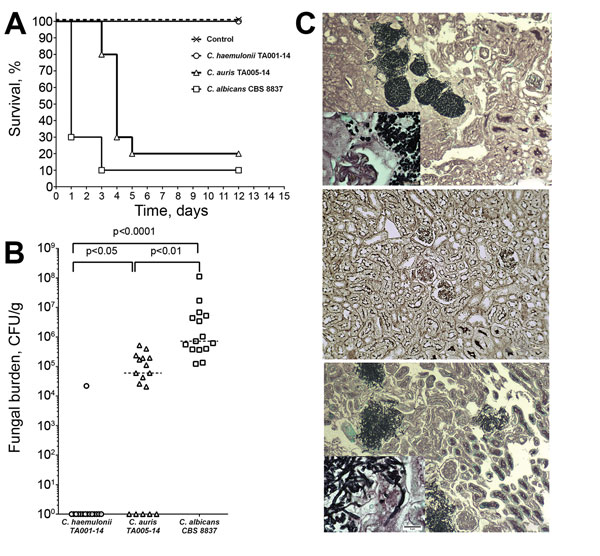
Figure 4. Differing virulence of Candida auris and C. haemulonii assessed in a mouse model of hematogenous disseminated candidiasis. Virulence was assessed in immunosuppressed BALB/c mice after intravenous injection of yeast cell suspension. A) Survival curves showing significantly shorter survival of mice infected with C. albicans than C. auris and no death among mice infected with C. haemulonii. B) Kidney fungal load (CFU per gram of tissue) shown to be significantly higher in mice infected with C. albicans than in those infected with C. auris, whereas no viable yeast was cultured from kidneys of mice infected with C. haemulonii. C) In mouse kidneys, C. auris cells formed aggregates and no hyphae (top) whereas C. albicans formed extensive tissue-invasive hyphae (bottom); C. haemulonii was not detected in tissue sections (middle). Grocott methenamine silver staining, original magnification ×100 for panels, ×400 for insets.

Main Article

Page created: January 12, 2017
Page updated: January 12, 2017
Page reviewed: January 12, 2017
The conclusions, findings, and opinions expressed by authors contributing to this journal do not necessarily reflect the official position of the U.S. Department of Health and Human Services, the Public Health Service, the Centers for Disease Control and Prevention, or the authors' affiliated institutions. Use of trade names is for identification only and does not imply endorsement by any of the groups named above.
file_external