Neurologic Complications of Influenza B Virus Infection in Adults, Romania
Corneliu P. Popescu

, Simin A. Florescu, Emilia Lupulescu, Mihaela Zaharia, Gratiela Tardei, Mihaela Lazar, Emanoil Ceausu, and Simona M. Ruta
Author affiliations: Carol Davila University of Medicine and Pharmacy, Bucharest, Romania (C.P. Popescu, S.A. Florescu, E. Ceausu, S.M. Ruta); Dr. Victor Babes Clinical Hospital of Infectious and Tropical Diseases, Bucharest (C.P. Popescu, S.A. Florescu, M. Zaharia, G. Tardei, E. Ceausu); European Society of Clinical Microbiology and Infection Study Group for Infectious Diseases of the Brain, Basel, Switzerland (C.P. Popescu, M. Zaharia); National Institute of Research Cantacuzino, Bucharest (E. Lupulescu, M. Lazar); Stefan S. Nicolau Institute of Virology, Bucharest (S.M. Ruta)
Main Article
Figure 1
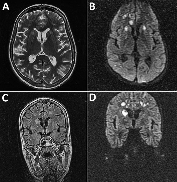
Figure 1. Magnetic resonance imaging of the brain of a 28-year-old woman (patient 1) who had neurologic complications of influenza B virus infection, Romania. A) Axial T2 image showing multiple areas of T2-associated hyperintense lesions with involvement of the genu corpus callosum, bilateral internal capsule, and several areas of white matter in the right frontal lobe, and more discreetly at the limit between the right parietal and occipital lobe. B) Axial diffusion-weighted image showing restricted diffusion associated with lesions. C) Coronal fluid–attenuated inversion recovery image showing multiple hyperintense lesions in the right caudate head and the cortical and deep white matter of the frontal lobes. D) Coronal diffusion-weighted image showing restricted diffusion associated with lesions.
Main Article
Page created: March 13, 2017
Page updated: March 13, 2017
Page reviewed: March 13, 2017
The conclusions, findings, and opinions expressed by authors contributing to this journal do not necessarily reflect the official position of the U.S. Department of Health and Human Services, the Public Health Service, the Centers for Disease Control and Prevention, or the authors' affiliated institutions. Use of trade names is for identification only and does not imply endorsement by any of the groups named above.