Volume 23, Number 6—June 2017
Dispatch
Severe Neurologic Disorders in 2 Fetuses with Zika Virus Infection, Colombia
Jorge Acosta-Reyes , Edgar Navarro, Maria José Herrera, Eloina Goenaga, Martha L. Ospina, Edgar Parra, Marcela Mercado, Pablo Chaparro, Mauricio Beltran, Maria Luz Gunturiz, Lissethe Pardo, Catalina Valencia, Sandra Huertas, Jorge Rodríguez, Germán Ruiz, Diana Valencia, Lisa B. Haddad, Sarah C. Tinker, Cynthia A. Moore, and Hernando Baquero
Figure 2
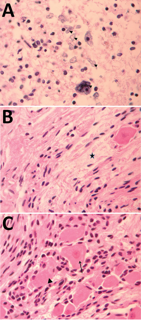
Figure 2. Pathology findings for case 2, involving a fetus examined after pregnancy termination who had severe neurologic defects attributed to maternal Zika virus infection, Colombia. A) Spinal cord slice showing neuropil disruption with multiple calcifications (arrowheads). B) Nerve showing disruptive changes of axons (Wallerian degeneration) (black star). C) Dorsal root ganglion showing spinal ganglion with satellitosis (arrow) and neuronophagia of ganglion cells (arrowhead). Hematoxylin and eosin staining; original magnification ×100.
Page created: May 16, 2017
Page updated: May 16, 2017
Page reviewed: May 16, 2017
The conclusions, findings, and opinions expressed by authors contributing to this journal do not necessarily reflect the official position of the U.S. Department of Health and Human Services, the Public Health Service, the Centers for Disease Control and Prevention, or the authors' affiliated institutions. Use of trade names is for identification only and does not imply endorsement by any of the groups named above.