Postmortem Findings for 7 Neonates with Congenital Zika Virus Infection
Anastácio Q. Sousa

, Diane I.M. Cavalcante, Luciano M. Franco, Fernanda M.C. Araújo, Emília T. Sousa, José Telmo Valença-Junior, Dionne B. Rolim, Maria E.L. Melo, Pedro D.T. Sindeaux, Marialva T.F. Araújo, Richard D. Pearson, Mary E. Wilson, and Margarida M.L. Pompeu
Author affiliations: Federal University of Ceará, Fortaleza, Brazil (A.Q. Sousa, D.I.M. Cavalcante, L.M. Franco, E.T. Sousa, J.T. Valença-Junior, P.D.T. Sindeaux, M.M.L. Pompeu); Serviço de Verificação de Óbitos-SVO, Fortaleza (L.M. Franco, E.T. Sousa, J.T. Valença-Junior); Ceará State Central Public Health Laboratory, Fortaleza (F.M.C. Araújo, M.E.L. Melo); University of Fortaleza, Fortaleza (D.B. Rolim); Ceará State Secretariat of Health, Fortaleza (D.B. Rolim); Evandro Chagas Institute, Belém, Brazil (M.T.F. Araújo); University of Virginia School of Medicine, Charlottesville, Virginia, USA (R.D. Pearson); University of California, San Francisco, California, USA (M.E. Wilson); Harvard T.H. Chan School of Public Health, Boston, Massachusetts, USA (M.E. Wilson)
Main Article
Figure 2
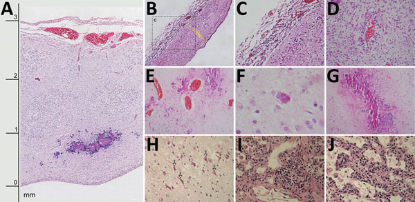
Figure 2. Histologic slides of tissues from 4 of 7 neonates who died of congenital Zika virus infection, Brazil. A) Neonate 1: severe cortical thinning (3 mm) with subventricular dystrophic calcification, reactive gliosis, and marked leptomeningeal congestion as well as marked depletion of neuronal precursors (original magnification ×10). B) Neonate 1: severe thinning of brain parenchyma (0.8 mm) with striking depletion of neuronal precursors (original magnification ×10). C) Neonate 1: lymphocytic leptomeningitis (enlargement of box in panel B; original magnification ×20). D) Neonate 6: white matter with lymphocytic perivascular cuffing and severe gliosis (original magnification ×40). E) Neonate 3: marked parenchymal vascular congestion and scattered coarse dystrophic calcification (original magnification ×20). F) Neonate 3: finely granular intracellular calcification (original magnification ×40). G) Neonate 7: band-like pattern of coarse dystrophic calcification at the junction of gray and white matter (original magnification ×10). H) Neonate 6: red neurons (arrows) in brain parenchyma (original magnification ×40). I) Neonate 1: focal interstitial lymphocytic pulmonary infiltrate (original magnification ×40). J) Neonate 1: expansion of alveolar septa with scattered lymphocytic and macrophage infiltrate (original magnification ×40).
Main Article
Page created: June 19, 2017
Page updated: June 19, 2017
Page reviewed: June 19, 2017
The conclusions, findings, and opinions expressed by authors contributing to this journal do not necessarily reflect the official position of the U.S. Department of Health and Human Services, the Public Health Service, the Centers for Disease Control and Prevention, or the authors' affiliated institutions. Use of trade names is for identification only and does not imply endorsement by any of the groups named above.