Skip directly to site content Skip directly to page options Skip directly to A-Z link Skip directly to A-Z link Skip directly to A-Z link
Volume 24, Number 1—January 2018
Research Letter

Yellow Fever Virus DNA in Urine and Semen of Convalescent Patient, Brazil

Carla M. Barbosa12, Nicholas Di Paola1, Marielton P. Cunha1, Mônica J. Rodrigues-Jesus, Danielle B. Araujo, Vanessa B. Silveira, Fabyano B. Leal, Flávio S. Mesquita, Viviane F. Botosso, Paolo M.A. Zanotto, Edison L. Durigon, Marcos V. Silva, and Danielle B.L. OliveiraComments to Author 
Author affiliations: University of São Paulo, São Paulo, Brazil (C.M. Barbosa, N. Di Paolo, M.P. Cunha, M.J. Rodrigues-Jesus, D.B. Araujo, V.B. Silveira, F.B. Leal, F.S. Mesquita, P.M.A. Zanotto, E.L. Durigon, D.B.L. Oliveira); Butantan Institute, São Paulo (V.F. Botosso); Institute of Infectology Emilio Ribas, São Paulo (M.V. Silva); Pontifical Catholic University, São Paulo (M.V. Silva)

Main Article

Figure

Clinical progression and detection of YFV in urine and semen of convalescent patient, Brazil. A) Platelet levels, clinical parameters and symptoms, and test results over a 128-day period after initial symptoms were observed. B) Maximum-likelihood tree (midpoint-rooted) inferred by using complete genomes of YFV to distinguish major virus genotypes; dashed box indicates South American clade strains, enlarged at top. Numbers near nodes indicate percent bootstrap values after 10,000 replicates for m

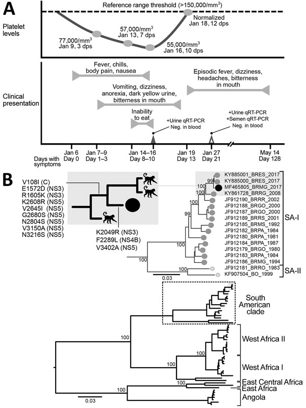
Figure. Clinical progression and detection of YFV RNA in urine and semen of convalescent patient, Brazil. A) Platelet levels, clinical parameters and symptoms, and test results over a 128-day period after initial symptoms were observed. B) Maximum-likelihood tree (midpoint-rooted) inferred by using complete genomes of YFV to distinguish major virus genotypes; dashed box indicates South American clade strains, enlarged at top. Numbers near nodes indicate percent bootstrap values after 10,000 replicates for major branches. Black circles indicate virus isolated in this study. Shaded boxes indicate monkey-derived virus sister taxa sampled during the same outbreak; inset at top left shows most parsimonious reconstructions of synapomorphic changes detected NS3, NS4B, and NS5 genes. GenBank accession number, geographic location code, and year of isolation are shown for virus isolates. Scale bars indicate nucleotide substitutions per site. C, capsid; dps, days postsymptom onset; Neg., negative; NS, nonstructural; qRT-PCR, quantitative reverse transcription PCR; SA, South America clade; YFV, yellow fever virus; +, positive.

Main Article

1These authors contributed equally to this article.

2Current affiliation: Pirbright Institute, Pirbright, UK.

Page created: December 19, 2017
Page updated: December 19, 2017
Page reviewed: December 19, 2017
The conclusions, findings, and opinions expressed by authors contributing to this journal do not necessarily reflect the official position of the U.S. Department of Health and Human Services, the Public Health Service, the Centers for Disease Control and Prevention, or the authors' affiliated institutions. Use of trade names is for identification only and does not imply endorsement by any of the groups named above.
file_external