Postmortem Findings in Patient with Guillain-Barré Syndrome and Zika Virus Infection
Emilio Dirlikov

, José V. Torres, Roosecelis Brasil Martines, Sarah Reagan-Steiner, George Venero Pérez, Aidsa Rivera, Chelsea Major, Desiree Matos, Jorge Muñoz-Jordan, Wun-Ju Shieh, Sherif Zaki, and Tyler M. Sharp
Author affiliations: Centers for Disease Control and Prevention, Atlanta, Georgia, USA (E. Dirlikov, R.B. Martines, S. Reagan-Steiner, C. Major, W.-J. Shieh, S.R. Zaki); Puerto Rico Institute of Forensic Sciences, San Juan, Puerto Rico, USA (J.V. Torres); Hermanos Meléndez Hospital, Bayamón, Puerto Rico, USA (G.V. Pérez); Centers for Disease Control and Prevention, San Juan (A. Rivera, C. Major, D. Matos, J. Muñoz-Jordan, T.M. Sharp); US Public Health Service Commissioned Corps, Rockville, Maryland, USA (T.M. Sharp)
Main Article
Figure 2
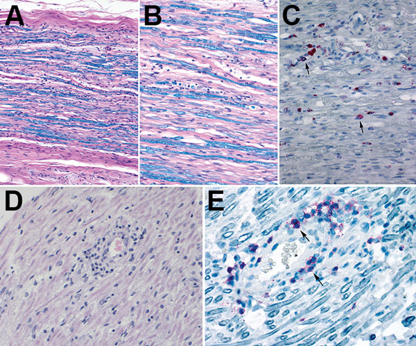
Figure 2. Histopathologic evaluation of tissue specimens collected postmortem from a patient with Guillain-Barré syndrome (acute demyelinating inflammatory polyneuropathy variant) and Zika virus infection, Puerto Rico, 2016. A, B) Luxol fast blue-periodic acid-Schiff myelin stain of sciatic nerves show patchy myelin loss and variable inflammation. Original magnification x10(A) and x20(B). C) Detection of CD68–positive cells (microphages) by immunohistochemistry (arrows) in sciatic nerve. Original magnification x20. D) Hematoxylin and eosin stain of cranial nerve IV shows perivascular lymphocytic infiltrate. Original magnification x20. E) Detection of T-lymphocytes by immunohistochemistry (arrows) in the same area where lymphocytic infiltrates were observed by hematoxylin and eosin stain. Original magnification x40.
Main Article
Page created: December 19, 2017
Page updated: December 19, 2017
Page reviewed: December 19, 2017
The conclusions, findings, and opinions expressed by authors contributing to this journal do not necessarily reflect the official position of the U.S. Department of Health and Human Services, the Public Health Service, the Centers for Disease Control and Prevention, or the authors' affiliated institutions. Use of trade names is for identification only and does not imply endorsement by any of the groups named above.