Volume 24, Number 11—November 2018
CME ACTIVITY - Synopsis
Rickettsia typhi as Cause of Fatal Encephalitic Typhus in Hospitalized Patients, Hamburg, Germany, 1940–1944
Jessica Rauch, Birgit Muntau, Petra Eggert, and Dennis Tappe
Figure 1
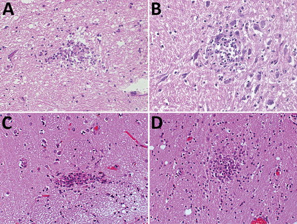
Figure 1. Hematoxylin and eosin staining of typical typhus nodules in brain of typhus patients during World War II, Hamburg, Germany, 1940–1944. Most nodules were found in the pons and medulla oblongata. A) Loose nodule. B) Spongy nodule amid large neuronal cells. C) Compact typhus nodule along longitudinal blood vessel. Note hyperemia of other blood vessels nearby. D) Another compact nodule with hyperemic blood vessels nearby. Original magnifications ×40.
Page created: October 15, 2018
Page updated: October 15, 2018
Page reviewed: October 15, 2018
The conclusions, findings, and opinions expressed by authors contributing to this journal do not necessarily reflect the official position of the U.S. Department of Health and Human Services, the Public Health Service, the Centers for Disease Control and Prevention, or the authors' affiliated institutions. Use of trade names is for identification only and does not imply endorsement by any of the groups named above.