Volume 24, Number 12—December 2018
Research
Novel Type of Chronic Wasting Disease Detected in Moose (Alces alces), Norway
Laura Pirisinu, Linh Tran, Barbara Chiappini, Ilaria Vanni, Michele A. Di Bari, Gabriele Vaccari, Turid Vikøren, Knut Ivar Madslien, Jørn Våge, Terry Spraker, Gordon Mitchell, Aru Balachandran, Thierry Baron, Cristina Casalone, Christer M. Rolandsen, Knut H. Røed, Umberto Agrimi, Romolo Nonno, and Sylvie L. Benestad
Figure 1
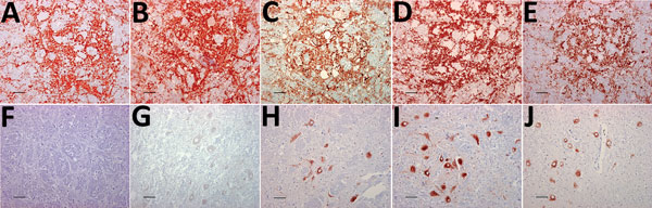
Figure 1. Immunohistochemical detection of disease-associated prion protein in brain sections at the level of the obex in cervids with chronic wasting disease, Norway. A–E) Reindeer; F–J) moose. mAbs used were 12B2 (A, F), 9A2 (B, G), L42 (C, H), SAF 84 (D, I), and F99/97.6 (E, J). Staining obtained in the reindeer tissues is similar regardless of mAbs used (A–E). Conversely, for moose tissues, the staining was primarily observed intraneuronally with L42, SAF84, and F99/97.6 (H–J) but was not observed using the more N-terminal mAbs 12B2 and 9A2 (F, G). Scale bars indicate 40 µm. mAbs, monoclonal antibodies.
Page created: November 20, 2018
Page updated: November 20, 2018
Page reviewed: November 20, 2018
The conclusions, findings, and opinions expressed by authors contributing to this journal do not necessarily reflect the official position of the U.S. Department of Health and Human Services, the Public Health Service, the Centers for Disease Control and Prevention, or the authors' affiliated institutions. Use of trade names is for identification only and does not imply endorsement by any of the groups named above.