Volume 24, Number 2—February 2018
Research Letter
Cronobacter sakazakii Infection from Expressed Breast Milk, Australia
Rowena McMullan, Vidthiya Menon, Alicia G. Beukers , Slade O. Jensen, Sebastiaan J. van Hal, and Rebecca Davis
Figure
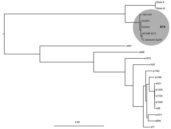
Figure. Maximum-likelihood phylogeny of Cronobacter isolates cultured from the blood of an infant (Ck0001) and the mother’s expressed breast milk (Ck0002) with C. sakazakii Sp291 as reference. Shaded circle highlights the clustering of sequence type 4 isolates. Scale bar indicates nucleotide substitutions per site. Methods for culturing isolates described in Technical Appendix. ST, sequence type.
Page created: January 17, 2018
Page updated: January 17, 2018
Page reviewed: January 17, 2018
The conclusions, findings, and opinions expressed by authors contributing to this journal do not necessarily reflect the official position of the U.S. Department of Health and Human Services, the Public Health Service, the Centers for Disease Control and Prevention, or the authors' affiliated institutions. Use of trade names is for identification only and does not imply endorsement by any of the groups named above.