Skip directly to site content Skip directly to page options Skip directly to A-Z link Skip directly to A-Z link Skip directly to A-Z link
Volume 24, Number 6—June 2018
Research

Prion Disease in Dromedary Camels, Algeria

Baaissa Babelhadj, Michele Angelo Di Bari, Laura Pirisinu, Barbara Chiappini, Semir Bechir Suheil Gaouar, Geraldina Riccardi, Stefano Marcon, Umberto Agrimi, Romolo Nonno, and Gabriele VaccariComments to Author 
Author affiliations: Ecole Normale Superieure Ouargla Laboratoire de Protection des Écosystèmes en Zones Arides et Semi Arides University Kasdi Merbah Ouargla, Ouargla, Algeria (B. Babelhadj); Istituto Superiore di Sanità Department of Food Safety, Nutrition and Veterinary Public Health, Rome, Italy (M.A. Di Bari, L. Pirisinu, B. Chiappini, G. Riccardi, S. Marcon, U. Agrimi, R. Nonno, G. Vaccari); Laboratoire de Physiopathologie et Biochimie de la Nutrition University Abou Bekr Bélkaid, Tlemcen, Algeria (S.B.S. Gaouar)

Main Article

Figure 1

Hematoxylin and eosin staining (A, B), immunohistochemistry (C–O), and paraffin-embedded tissue blot analysis (P–S) of brains of dromedary camels brought for slaughter to the Ouargla abattoir, Algeria, 2015–2016. Spongiform change of neuropil, gliosis, and neuronal loss in thalamus (A) and intraneuronal vacuolation in pons (B) (scale bar = 50 μm). Immunohistochemistry for prion protein (PrPSc) with L42 monoclonal antibody evidenced dense synaptic/punctate deposition in thalamus (C) and intraneur

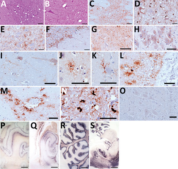
Figure 1. Hematoxylin and eosin staining (A, B), immunohistochemistry (C–O), and paraffin-embedded tissue blot analysis (P–S) of brains of dromedary camels brought for slaughter to the Ouargla abattoir, Algeria, 2016–2017. Spongiform change of neuropil, gliosis, and neuronal loss in thalamus (A) and intraneuronal vacuolation in pons (B) (scale bar = 50 μm). Immunohistochemistry for prion protein (PrPSc) with L42 monoclonal antibody evidenced dense synaptic/punctate deposition in thalamus (C) and intraneuronal and extraneuronal PrPSc deposits in pons (D), accompanied by spongiform change. Perineuronal, diffused in neuropil and glial-associated PrPSc staining were also observed in the nucleus of the solitary tract (E) and cerebellum (F), which showed rare vacuoles (scale bars = 50 μm). Immunohistochemical analysis performed on brains of symptomatic dromedaries revealed several PrPSc deposition patterns, such as synaptic/punctate pattern diffused in the neuropil (G); intraneuronal deposition in pyramidal cells of hippocampus (H); perineuronal and linear staining in frontal cortex (I); intraglial PrPSc deposition (J–L); perivascular deposition (M); atypical intracellular PrPSc deposition pattern in pons (N). PrPSc was absent in asymptomatic dromedary used as negative control (O) (scale bars = 50 μm). PrPSc distribution, by paraffin-embedded tissue blot analysis, was observed in several brain areas, such as prefrontal cortex (P), hippocampus (Q), cerebellum (R), and a sagittal section of pons (S) (scale bar = 3 mm).

Main Article

Page created: May 17, 2018
Page updated: May 17, 2018
Page reviewed: May 17, 2018
The conclusions, findings, and opinions expressed by authors contributing to this journal do not necessarily reflect the official position of the U.S. Department of Health and Human Services, the Public Health Service, the Centers for Disease Control and Prevention, or the authors' affiliated institutions. Use of trade names is for identification only and does not imply endorsement by any of the groups named above.
file_external