Skip directly to site content Skip directly to page options Skip directly to A-Z link Skip directly to A-Z link Skip directly to A-Z link
Volume 24, Number 6—June 2018
Research

Prion Disease in Dromedary Camels, Algeria

Baaissa Babelhadj, Michele Angelo Di Bari, Laura Pirisinu, Barbara Chiappini, Semir Bechir Suheil Gaouar, Geraldina Riccardi, Stefano Marcon, Umberto Agrimi, Romolo Nonno, and Gabriele VaccariComments to Author 
Author affiliations: Ecole Normale Superieure Ouargla Laboratoire de Protection des Écosystèmes en Zones Arides et Semi Arides University Kasdi Merbah Ouargla, Ouargla, Algeria (B. Babelhadj); Istituto Superiore di Sanità Department of Food Safety, Nutrition and Veterinary Public Health, Rome, Italy (M.A. Di Bari, L. Pirisinu, B. Chiappini, G. Riccardi, S. Marcon, U. Agrimi, R. Nonno, G. Vaccari); Laboratoire de Physiopathologie et Biochimie de la Nutrition University Abou Bekr Bélkaid, Tlemcen, Algeria (S.B.S. Gaouar)

Main Article

Figure 3

Western blot analysis of dromedary prion protein (PrPSc). A) Western blot analysis of proteinase K (PK)–treated PrPSc in brain homogenates from dromedary camels with neurologic symptoms (#4 and #8), Ouargla abattoir, Algeria. A sample of sheep scrapie was loaded as control (indicated as C+). Membranes were probed with L42 (left) and 12B2 monoclonal antibody (mAb) (right). Molecular weights (kDa) are indicated on the left. Tissue equivalents loaded per lane were 2 mg for camel samples and 0.1 mg

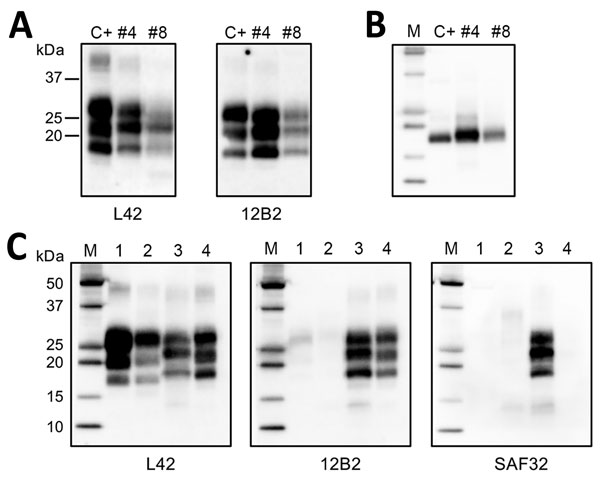
Figure 3. Western blot analysis of protein-resistant core (PrPres) of pathological dromedary prion protein. A) Western blot analysis of proteinase K (PK)–treated PrPSc in brain homogenates from dromedary camels with neurologic symptoms (nos. 4 and 8), Algeria. A sample of sheep scrapie was loaded as control (indicated as C+). Membranes were probed with L42 (left) and 12B2 monoclonal antibody (mAb) (right). Molecular weights (kDa) are indicated on the left. Tissue equivalents loaded per lane were 2 mg for camel samples and 0.1 mg for sheep scrapie. B) Samples after deglycosylation. Membrane was probed with L42 mAb. C) Comparison of dromedary PrPres (from camel no. 4) with sheep bovine spongiform encephalopathy (BSE), bovine BSE, and sheep scrapie samples by ISS (Istituto Superiore di Sanità) discriminatory Western blot (17). Tissue equivalents loaded per lane were 2 mg for dromedary camel and bovine samples and 0.1 mg for sheep samples. In each blot, samples were loaded as follows: lane 1, ovine BSE; lane 2, bovine BSE; lane 3, dromedary camel no. 4; lane 4, ovine scrapie. Membranes were probed with L42, 12B2, and SAF32 mAbs, as indicated. For the analyses in panels B and C, protein standards were loaded and are indicated as M.

Main Article

Page created: May 17, 2018
Page updated: May 17, 2018
Page reviewed: May 17, 2018
The conclusions, findings, and opinions expressed by authors contributing to this journal do not necessarily reflect the official position of the U.S. Department of Health and Human Services, the Public Health Service, the Centers for Disease Control and Prevention, or the authors' affiliated institutions. Use of trade names is for identification only and does not imply endorsement by any of the groups named above.
file_external