Skip directly to site content Skip directly to page options Skip directly to A-Z link Skip directly to A-Z link Skip directly to A-Z link
Volume 24, Number 6—June 2018
Research

Influenza D Virus Infection in Feral Swine Populations, United States

Lucas Ferguson1, Kaijian Luo1, Alicia K. Olivier, Fred L. CunninghamComments to Author , Sherry Blackmon, Katie Hanson-Dorr, Hailiang Sun2, John Baroch, Mark W. Lutman, Bianca Quade, William Epperson, Richard Webby, Thomas J. DeLiberto, and Xiu-Feng WanComments to Author 
Author affiliations: Mississippi State University, Starkville, Mississippi, USA (L. Ferguson, K. Luo, A.K. Olivier, S. Blackmon, H. Sun, B. Quade, W. Epperson, X.-F. Wan); South China Agricultural University, Guangzhou, China (K. Luo); US Department of Agriculture, Starkville (F.L. Cunningham, K. Hanson-Dorr); US Department of Agriculture, Fort Collins, Colorado, USA (J. Baroch, M.W. Lutman, T.J. DeLiberto); St. Jude Children’s Research Hospital, Memphis, Tennessee, USA (R. Webby)

Main Article

Figure 3

Influenza D viral titers in feral swine tissues. Feral swine were inoculated intranasally with 106 TCID50 of influenza D/bovine/C00046N/Mississippi/2014 virus or sterile phosphate-buffered saline (controls). At 3, 5, 7, 9, and 11 days postinoculation, they were humanely euthanized, and the following tissues were collected: turbinate; soft palate; upper, middle, and lower trachea; bronchus; left and right caudal lung; left and right medial lung; left and right cranial lung; and right accessory lu

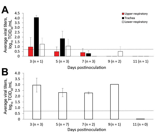
Figure 3. Influenza D viral titers in feral swine tissues. Feral swine were inoculated intranasally with 106 TCID50 of influenza D/bovine/C00046N/Mississippi/2014 virus or sterile phosphate-buffered saline (controls). At 3, 5, 7, 9, and 11 days postinoculation, they were humanely euthanized, and the following tissues were collected: turbinate; soft palate; upper, middle, and lower trachea; bronchus; left and right caudal lung, left and right medial lung, left and right cranial lung; and right accessory lung. A) The tissues were grouped as upper respiratory tract (turbinate and soft palate), trachea (upper, middle, and lower trachea, and bronchus), and lower respiratory tract (left and right caudal lung; left and right medial lung; and left and right cranial lung). B) All lung tissue sections at each time point that were influenza D virus–positive by TCID50 titration in HRT-18G cells were averaged and plotted for each day postinoculation; day 9 has no error bars because only 1 positive tissue sample was found. Dashed lines indicate the lower limit of detection, which was 100.699 TCID50/mL. Error bars indicate SE. Numbers in parentheses indicate number of animals used in the analyses. TCID50, 50% tissue culture infective dose.

Main Article

1These authors contributed equally to this article.

2Current affiliation: South China Agricultural University, Guangzhou, China.

Page created: May 17, 2018
Page updated: May 17, 2018
Page reviewed: May 17, 2018
The conclusions, findings, and opinions expressed by authors contributing to this journal do not necessarily reflect the official position of the U.S. Department of Health and Human Services, the Public Health Service, the Centers for Disease Control and Prevention, or the authors' affiliated institutions. Use of trade names is for identification only and does not imply endorsement by any of the groups named above.
file_external