Skip directly to site content Skip directly to page options Skip directly to A-Z link Skip directly to A-Z link Skip directly to A-Z link
Volume 24, Number 8—August 2018
Dispatch

Invasive Colonic Entamoebiasis in Wild Cane Toads, Australia

Cathy M. Shilton, Jan Šlapeta, Richard Shine, and Gregory P. BrownComments to Author 
Author affiliations: Northern Territory Department of Primary Industry and Resources, Darwin, Northern Territory, Australia (C.M. Shilton); University of Sydney, Sydney, New South Wales, Australia (J. Šlapeta, R. Shine, G.P. Brown)

Main Article

Figure 1

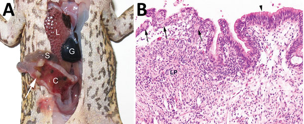
Invasive colonic entamoebiasis in wild cane toads (Rhinella marina), tropical Australia, 2014–2015. A) Toad with severe colonic amebiasis. The colon (C) has been opened to show intraluminal hemorrhagic content and blood clots. There is segmental full-thickness necrosis of the colon wall (white arrow). Lung (L), small intestine (S), and gall bladder (G) are annotated for perspective. B) Photomicrograph of colonic amebiasis. The affected segment of mucosal epithelium, which contains several amebae

Figure 1. Invasive colonic entamoebiasis in wild cane toads (Rhinella marina), tropical Australia, 2014–2015. A) Toad with severe colonic amebiasis. The colon (C) has been opened to show intraluminal hemorrhagic content and blood clots. There is segmental full-thickness necrosis of the colon wall (white arrow). Lung (L), small intestine (S), and gall bladder (G) are annotated for perspective. B) Photomicrograph of colonic amebiasis. The affected segment of mucosal epithelium, which contains several amebae (arrows) is jumbled and sloughing from the underlying lamina propria (LP). Relatively normal colonic epithelium is present at right (arrowhead). There is lymphohistiocytic and granulocytic infiltration of the lamina propria underlying the affected epithelium. Hematoxylin and eosin stain. Original magnification ×200.

Main Article

Page created: July 18, 2018
Page updated: July 18, 2018
Page reviewed: July 18, 2018
The conclusions, findings, and opinions expressed by authors contributing to this journal do not necessarily reflect the official position of the U.S. Department of Health and Human Services, the Public Health Service, the Centers for Disease Control and Prevention, or the authors' affiliated institutions. Use of trade names is for identification only and does not imply endorsement by any of the groups named above.
file_external