Fatal Tickborne Phlebovirus Infection in Captive Cheetahs, Japan
Keita Matsuno

, Noriyuki Nonoue, Ayako Noda, Nodoka Kasajima, Keita Noguchi, Ai Takano, Hiroshi Shimoda, Yasuko Orba, Mieko Muramatsu, Yoshihiro Sakoda, Ayato Takada, Shinji Minami, Yumi Une, Shigeru Morikawa, and Ken Maeda
Author affiliations: Hokkaido University, Sapporo, Japan (K. Matsuno, Y. Sakoda, A. Takada); Hiroshima City Asa Zoological Park, Hiroshima, Japan (N. Nonoue, A. Noda, S. Minami); Hokkaido University Research Center for Zoonosis Control, Sapporo (N. Kasajima, Y. Orba, M. Muramatsu, A. Takada); Yamaguchi University, Yamaguchi, Japan (K. Noguchi, A. Takano, H. Shimoda, K. Maeda); Okayama University of Science, Imabari, Japan (Y. Une); National Institute of Infectious Diseases, Tokyo, Japan (S. Morikawa)
Main Article
Figure 1
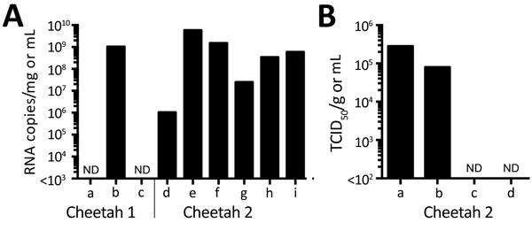
Figure 1. Detection of severe fever with thrombocytopenia syndrome virus (SFTSV) in samples from 2 cheetahs, Japan, 2017. A) RNA was extracted from tissues, plasma, and serum and subjected to quantitative reverse transcription PCR (RT-PCR). The amounts of SFTSV RNA were quantified, with a reference, as RNA copies/mg for tissues and RNA copies/mL for plasma and serum. The mean of duplicate results is shown in the graph. a, plasma; b, popliteal lymph node (left); c, serum; d, brain; e, salivary gland; f, spleen; g, mesentric lymph node; h, popliteal lymph node (left); i, popliteal lymph node (right). B) The TCID50 of salivary gland (per mg) and swab specimens (per mL) for cheetah 2 was determined using Huh-7 cells. Virus proteins were detected by an immunofluorescence assay with an anti-SFTSV N monoclonal antibody. a, salivary gland; b, oral swab sample; c, nasal swab sample; d, rectal swab sample. ND, not done; TCID50, 50% tissue culture infectious dose.
Main Article
Page created: August 14, 2018
Page updated: August 14, 2018
Page reviewed: August 14, 2018
The conclusions, findings, and opinions expressed by authors contributing to this journal do not necessarily reflect the official position of the U.S. Department of Health and Human Services, the Public Health Service, the Centers for Disease Control and Prevention, or the authors' affiliated institutions. Use of trade names is for identification only and does not imply endorsement by any of the groups named above.