Volume 25, Number 1—January 2019
CME ACTIVITY - Synopsis
Enterovirus A71 Infection and Neurologic Disease, Madrid, Spain, 2016
Carmen Niño Taravilla1 , Isabel Pérez-Sebastián1, Alberto García Salido, Claudia Varela Serrano, Verónica Cantarín Extremera, Anna Duat Rodríguez, Laura López Marín, Mercedes Alonso Sanz, Olga María Suárez Traba, and Ana Serrano González
Figure 2
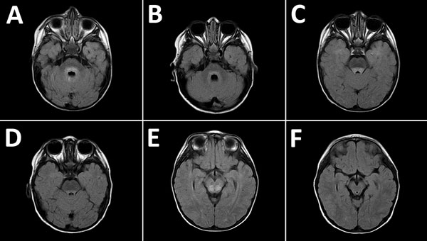
Figure 2. Magnetic resonance images of the brain of a 2-year-old boy with enterovirus meningoencephalitis. A–C) Brain at time of diagnosis. FLAIR sequences show hyperintense lesions around ventricle IV (A), posterior region of the pons (B), and posterior region of the mesencephalon (C). D–F) Control images of cerebrum 6 months after diagnosis. FLAIR sequences show slight hyperintensity of signal around ventricle IV, lower than in the initial study (D), and complete resolution of lesions in the posterior region of the pons (E) and mesencephalon (F).
1These authors contributed equally to this article.
Page created: December 13, 2018
Page updated: December 13, 2018
Page reviewed: December 13, 2018
The conclusions, findings, and opinions expressed by authors contributing to this journal do not necessarily reflect the official position of the U.S. Department of Health and Human Services, the Public Health Service, the Centers for Disease Control and Prevention, or the authors' affiliated institutions. Use of trade names is for identification only and does not imply endorsement by any of the groups named above.