Volume 25, Number 1—January 2019
CME ACTIVITY - Synopsis
Enterovirus A71 Infection and Neurologic Disease, Madrid, Spain, 2016
Carmen Niño Taravilla1 , Isabel Pérez-Sebastián1, Alberto García Salido, Claudia Varela Serrano, Verónica Cantarín Extremera, Anna Duat Rodríguez, Laura López Marín, Mercedes Alonso Sanz, Olga María Suárez Traba, and Ana Serrano González
Figure 3
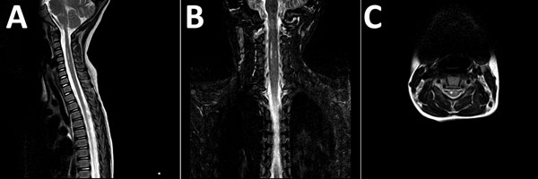
Figure 3. Magnetic resonance images of brain of a 3-year-old girl with enterovirus encephalomyelitis (paresis of the right upper limb). A) Image of the cervical spine: sagittal T2 sequence; B) short tau inversion recovery (STIR) coronal sequence; C) T2 axial sequence. Hyperintense filiform lesions in the anterolateral regions of the spinal cord (C3–C5), predominantly right, are suggestive of myelitis.
1These authors contributed equally to this article.
Page created: December 13, 2018
Page updated: December 13, 2018
Page reviewed: December 13, 2018
The conclusions, findings, and opinions expressed by authors contributing to this journal do not necessarily reflect the official position of the U.S. Department of Health and Human Services, the Public Health Service, the Centers for Disease Control and Prevention, or the authors' affiliated institutions. Use of trade names is for identification only and does not imply endorsement by any of the groups named above.